
Jason Fink, BS OMS 20271; Arash Badragheh, BS OMS 20271; Nikhil Sai, MS OMS 20281; Tobi Salako, DO, Intern Physician2; Bradley A. Fink, DO, Orthopedic Surgeon2
1Philadelphia College of Osteopathic Medicine
2TrinityHealth Mid-Atlantic Nazareth Hospital
DOI: 10.70709/feguw7qn3z
Abstract
The authors present a case involving a boxer fracture with an atypical presentation and course. Initial evaluation and skin inspection led to the impression of a “routine” closed boxer fracture that we commonly see in our clinic. During treatment, however, it became apparent that the fracture was open and complicated by metacarpophalangeal (MCP) septic arthritis. Surgical intervention resulted in infection eradication and successful fracture union. Open boxer fractures are uncommonly seen and rarely reported. Signs of an open boxer fracture may be easily overlooked, and the consequences can be devastating. Doctors may miss a deeper injury if not making a conscious effort to consider it in every patient. It is recommended that all boxer fractures be considered a fight bite injury and thus an open fracture during initial and follow-up evaluations. This approach should be taken by any physician that manages acute clenched fist fractures to the hand.
Keywords: Open boxer fracture, fight bite, metacarpophalangeal septic arthritis
Introduction
A boxer fracture consists of fracture of the 5th metacarpal neck that occurs when a clenched fist impacts a person or object. Although most are closed, an open fracture may not be obvious. A skin breach may go unnoticeable to the patient and undetectable to the physician. Also, it may appear as an insignificant, superficial wound to the patient and they may omit it from their history. Additionally, a skin breach may not be visible on examination by the time the patient presents to the physician. A neglected or misdiagnosed open boxer fracture may lead to serious complications and should be considered on the initial presentation, and during each follow up visit.(1)
Case Report
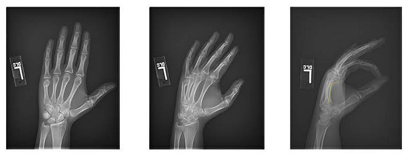
A 24-year-old female presented with mild dorsal left-hand pain localized to the 4th and 5th MCP joints after punching a door two days prior. She denied laceration and drug abuse. She had relief with Motrin and a volar splint. According to the emergency room record, the patient stated that the injury occurred after punching someone in the face. Past medical history was negative. Examination revealed dorsal hand swelling and ecchymosis without deformity, erythema, cellulitis, subungual hematoma, laceration, drainage, localization or fluctuance. The emergency room record also documented intact skin. X-rays demonstrated a 43-degree angulated fifth metacarpal neck fracture. A brace was applied. One week later, the Motrin was no longer relieving her pain, scissoring was noted and repeat x-rays confirmed 62-degrees of fracture angulation (Fig.1).
The following week, the patient was scheduled for open reduction internal fixation. Two grams of cefazolin were given intravenously and upon dorsal MCP arthrotomy, purulent material was released. The joint purulence was continuous with the fracture site, confirming joint sepsis and acute osteomyelitis of the 5th metacarpal neck fracture. An intraoperative gram stain and culture were obtained, the joint and fracture site were lavaged with saline, the fracture was reduced using the Jahss maneuver and the skin was loosely approximated to provide bone, extensor tendon and joint coverage while allowing for drainage. An ulnar gutter splint was applied. The gram stain revealed moderate gram-positive cocci. This was likely Staphylococcus Aureus, the most common fight bite infection. Sero markers for infection were not obtained because the diagnosis was confirmed and the patient was going to be monitored clinically and radiographically as she was young and healthy and the infection was acute. Infectious disease was consulted to whom the patient confirmed that there was no skin laceration after the index injury. The patient was given a dose of vancomycin and discharged to receive intravenous dalbavancin as an outpatient the following day. Dalbavancin was chosen because it provides the same coverage as vancomycin in a one-time dose while avoiding the need of a PICC line for 4 weeks and the inability to perform self-infusions due to the hand injury. After two weeks, the splint was discontinued and physical therapy was initiated. Culture was negative, likely due to the preoperative cefazolin. One week later, there was no swelling, deformity, skin changes, fracture tenderness, drainage, loculation or fluctuance. Hand cascade was normal, the wound was healed, and full, painless active range of motion was obtained. There was no sequela at 2 years post injury.
Discussion
Boxer fractures are the most common metacarpal fracture, accounting for 20% of all hand fractures.(2,3,4,5) While the majority of boxer fractures are closed, open boxer fractures rarely occur and their incidence is unknown. This is partly because some patients don’t disclose an accurate history.(6,7) For example, the presence of a laceration is underreported by patients who feel that the injury is minor and don’t appreciate the severity of the wound and injury. Additionally, some patients may be embarrassed about the injury mechanism and/or fear hospitals and legal repercussion. (8,9) Clenched fist injuries, therefore, often present late and may result in complications.(10) Such complications include; septic arthritis, septic tenosynovitis, osteomyelitis, amputation and death.(7,8,9,10,11)
There are more data regarding fight bite injuries than open boxer fractures. A fight bite is a human bite wound to the hand that occurs when a clenched fist contacts a tooth. Unlike boxer fractures, fight bite injuries are commonly seen over the third and fourth MCP joints.(1,7,8) The infection rate following fight bite wounds is 10% to 50%. (1,6,7,8) Thirty percent of these cases are septic arthritis, 40% are osteomyelitis and 25% are tenosynovitis.(7) One study showed an 18% amputation rate due to osteomyelitis or chronic tenosynovitis.(9) In another study of hand osteomyelitis, the amputation rate was 39% which climbed to 86% when there was a 6 month delay in diagnosis.(12) S. Aureus is the most common acute fight bite infection while Eikenella Corrodens is associated with delayed presentation.(10) It is therefore imperative that a careful history and examination be pursued with every boxer fracture. (3,11,13) A fight bite is deceptive and dangerous and may appear innocuous causing the examiner to miss or not appreciate the seriousness of the wound.(9,13) A small breach in the skin may not be apparent on examination and therefore it is important to ask patients with boxer fractures if they had noticed a laceration or bleeding around the knuckle at the time of injury. The examiner should consider all wounds located to the dorsal MCP joint to be a fight bite.(6,7) Wounds may be subtle and appear as an abrasion or superficial laceration.(6) Many present as a puncture wound that does not appear to penetrate deep.(9) Small lacerations may seem minor and without a breach in the capsule or the extensor tendon.(1) It is best to examine the dorsal skin at the MCP joint for abrasions and lacerations through a full range of motion, especially in a clenched fist position.(4,6,7) Teeth produce a 3-5mm laceration over the MCP joint.(6) A tooth mark on the skin may be associated with a deeper infection.(10) Wounds or teeth marks near the MCP joint after a clenched fist injury are often misleading and should be cautiously evaluated.(1,9,10)
Lacerations occur with a flexed MCP joint followed by extension allowing the extensor tendon to drag bacteria proximally, creating a deep bacterial inoculation. The skin and capsule also retract proximally closing off the wound.(6,8) Typically, the hand is examined in extension and injury to the deeper structures, which have retracted proximally, may go unnoticed.(9) Subtle lacerations may be missed in extension as the skin is not taut. There is a likelihood of missing lacerations and deeper injuries when the examination is done in extension.(10) Wounds must be explored for tendon and capsule laceration.(6,8) Penetration of the MCP joint capsule occurs in 95% of cases when the skin is broken.(7) Occasionally, a chondral divot fracture may also occur.(8) While assessing x-rays for fracture, also look for tooth fragmentation.(7,10)
E. Corrodens and anaerobes are difficult to grow in vitro because they require optimal conditions to survive which may result in a false negative culture. Antibiotics should be given, without delay, even with negative cultures.(8,9) Broad-spectrum antibiotics, such as a loading dose of Unasyn followed by Augmentin, is recommended to cover a polymicrobial infection including; S. Aureus (most common), Streptococcus, Corynebacterium and E. Corrodens.(1,6,10,11) Tetanus immunization should be addressed because 50% of human bites transmit Clostridium Tetani.(6) Wound irrigation with dilute povidone-iodine or dilute hydrogen peroxide should be used.(8,9) Wounds should be left open.(9,10)
Fight bite infections can present 12 hours post injury and may initially go undetected. (7,8) It is critical to re-evaluate the patient within 48 hours to monitor for infection as early intervention prevents septic arthritis and osteomyelitis.(10) Infection should be considered if the patient’s pain level does not subside, or standard analgesics do not provide relief as seen in this case. Sinha, et al demonstrated a correlation between delayed treatment of 3 days and septic arthritis. Goon, et al found a 66% infection rate four days post fight bite, most of which involved the MCP joint or tendon.(8) Ten percent of patients with MCP pyogenic arthritis regain normal hand function.(6,7)
Hand anatomy and injury mechanism predispose people to open fracture and infection. (8) The 5th MCP joint is covered dorsally by thin skin while the extensor tendon and capsule are superficial.(6,8) The flexed position at the time of injury makes these structures taut and easily penetrable.(8) The articular cartilage, extensor tendon, joint capsule and synovium are relatively avascular and compartmentalized hampering the immune response and precludes drainage.(6,8) The joint space and synovial fluid provide a medium for bacterial growth.(8)
It is plausible that the progressive fracture displacement, during the course of treatment, was directly related to the infection and that such secondary displacement may be a sign of infection.
Conclusion
Closed boxer fractures are commonly encountered in the outpatient setting. An open injury may appear benign but become limb or life threatening. We present this case to make physicians and other healthcare workers aware of the subtle presentation of open boxer fractures and to offer an approach to identify them early. Upon presentation, our patient appeared to have a routine closed boxer fracture. During treatment, the diagnosis of open fracture with septic arthritis and osteomyelitis became apparent. Early arthrotomy, debridement, reduction and intravenous antibiotics resulted in eradication of the infection, union of the fracture and a functional hand. Healthcare workers should consider all boxer fractures as an open injury until proven otherwise during treatment. All patients with boxer fractures must be questioned about skin integrity at the time of injury and carefully examined for MCP wounds in the clenched fist position. If present, wounds should be considered penetrating and treated as such.(9) Secondary fracture displacement and pain that does not improve with time or analgesics should be re-evaluated for a possible deep infection. These suggestions will facilitate early detection of an open fracture so that appropriate and timely treatment may be incorporated to prevent chronic and potentially devastating sequela. While most of these injuries are closed, the examiner must maintain a high index of suspicion for an open fracture due to patient underreporting and their propensity for infection.(3,8,10)
Consent for publication
Informed consent was obtained from the patient who agreed to the publication of this case report and all information and data contained within.
References
- Sbai MA, Benzarti S, Boussen M, Maalla R. Teeth syndrome: diagnosis, complications and management. Pan Afr Med J. 2015 Sep 28;22:71. doi: 10.11604/pamj.2015.22.71.7313. PMID: 26834924; PMCID: PMC4725650.
- Luciani MA, Mayers Y, Warnick EP, Udoeyo IF, Klena JC, Grandizio LC. Trends in the Management of Fifth Metacarpal Neck Fractures. J Hand Surg Am. 2024 Apr;49(4):384.e1-384.e9. doi: 10.1016/j.jhsa.2022.08.008. Epub 2022 Oct 7. PMID: 36216683.
- Wormald J, Claireaux HA, Gardiner MD, Jain A, Furniss D, Costa ML. Management of extra-articular fractures of the fifth metacarpal: Operative vs. Non-opeRaTive TrEatment (FORTE) – A systematic review and meta-analysis. JPRAS Open. 2019 Feb 15;20:59-71. doi: 10.1016/j.jpra.2019.02.001. PMID: 32158872; PMCID: PMC7061598
- Hussain MH, Ghaffar A, Choudry Q, Iqbal Z, Khan MN. Management of Fifth Metacarpal Neck Fracture (Boxer’s Fracture): A Literature Review. Cureus. 2020 Jul 28;12(7):e9442. doi: 10.7759/cureus.9442. PMID: 32864266; PMCID: PMC7451089.
- Lambi AG, Rowland RJ, Brady NW, Rodriguez DE, Mercer DM. Metacarpal fractures. Journal of Hand Surgery (European Volume). 2023;48(2_suppl):42S-50S. doi:10.1177/17531934231184119
- Perron AD, Miller MD, Brady WJ. Orthopedic pitfalls in the ED: fight bite. Am J Emerg Med. 2002 Mar;20(2):114-7. doi: 10.1053/ajem.2002.31146. PMID: 11880877.
- Suzanne Alton, Nikoletta Carayannopoulos,Hand Injuries: Boxer Fractures and Fight Bites, The Journal for Nurse Practitioners, Volume 15, Issue 5, 2019, Pages 334-338, ISSN 1555-4155, https://doi.org/10.1016/j.nurpra.2018.12.017. (https://www.sciencedirect.com/science/article/pii/S1555415518307803)
- Goon PK, Mahmoud M, Rajaratnam V. Hand Trauma Pitfalls: A Retrospective Study of Fight Bites. Eur J Trauma Emerg Surg. 2008 Apr;34(2):135-40. doi: 10.1007/s00068-007-6183-9. Epub 2007 Oct 11. PMID: 26815618.
- Mennen U, Howells CJ. Human fight-bite injuries of the hand. A study of 100 cases within 18 months. J Hand Surg Br. 1991 Nov;16(4):431-5. doi: 10.1016/0266-7681(91)90020-o. PMID: 1779160.
- Raval P, Khan W, Haddad B, Mahapatra AN. Bite injuries to the hand – review of the literature. Open Orthop J. 2014 Jun 27;8:204-8. doi: 10.2174/1874325001408010204. PMID: 25097675; PMCID: PMC4112338.
- Harper CM, Dowlatshahi AS, Rozental TD. Challenging Dogma: Optimal Treatment of the “Fight Bite”. Hand (N Y). 2020 Sep;15(5):647-650. doi: 10.1177/1558944719831238. Epub 2019 Feb 27. PMID: 30808240; PMCID: PMC7543217.
- Reilly KE, Linz JC, Stern PJ, Giza E, Wyrick JD. Osteomyelitis of the tubular bones of the hand. J Hand Surg Am. 1997 Jul;22(4):644-9. doi: 10.1016/ S0363-5023(97)80122-0. PMID: 9260620.
- Meals C, Meals R. Hand fractures: a review of current treatment strategies. J Hand Surg Am. 2013 May;38(5):1021-31; quiz 1031. doi: 10.1016/ j.jhsa.2013.02.017. PMID: 23618458.
- Sinha M, Jain S, Woods DA. Septic Arthritis of the Small Joints of the Hand. Journal of Hand Surgery. 2006;31(6):665-672. doi:10.1016/J.JHSB.2006.08.011
