
Anthony Carano B.S., O.M.S-III1; Catherine Magro B.S., O.M.S-III; Jordan Read DO, ATC2; , Gregory Hill DO, FAOAO2
1Ohio University Heritage College of Osteopathic Medicine
2Western Reserve Hospital
Abstract
Introduction
Tumoral Calcinosis (TC) is a rare and severe complication of hemodialysis characterized by calcium salt deposits in periarticular soft tissue, often involving the hips, elbows, shoulders, and scapulae. Finger involvement, especially isolated, is exceedingly rare.
Case Report
We report a case of Secondary Tumoral Calcinosis in a 54-year-old male with chronic kidney disease on hemodialysis involving multiple areas of the hands, including the right dorsal ring finger, left volar middle finger, and left volar ring finger. We collected a thorough medical history, including laboratory values, x-ray imaging, and pathologic examination to determine this precise diagnosis. Given the sequelae of this disease experienced by this patient, we performed surgical excision of the largest calcification for symptomatic treatment.
Conclusions
TC may be rare but can affect end stage renal disease patients on hemodialysis and should be suspected even when presenting in uncommon locations. Physical exam and imaging are imperative in diagnosis, and surgical excision remains the mainstay treatment for symptomatic patients.
Keywords: Calcification, Tumoral Calcinosis, Hemodialysis, Hand, Digits
Introduction
Tumoral Calcinosis (TC) is a benign condition described as periarticular tumor-like calcium deposits in soft tissue, bone, and periarticular spaces (1). The term has been used loosely over the years to describe any calcification of periarticular soft tissue. TC can be primary, as in the case of familial or sporadic subtypes, but it is more often seen as a secondary manifestation of other diseases or conditions. Most notably chronic kidney disease (CKD), affecting approximately 1.6% of dialysis patients (1,2). There is a predilection for these calcified lesions to occur in the hips, elbows, shoulders, and scapulae (1). A very rare location for TC to present is in the hands (3). Whereas primary TC classically presents as rubbery, nontender swellings and nodules in affected areas, patients with secondary TC more commonly report the addition of pain and decreased mobility (2,4). X-ray imaging reveals dense masses of calcification that may resemble a “chicken wire” appearance (1).
This case report highlights a case of secondary TC of the hands in a patient requiring hemodialysis. We searched for similar cases in the PubMed database using the “Advanced Search” option and selecting “title” in the advanced search builder. We utilized searches with the following search term combinations: “tumoral calcinosis”, “hand”, “dialysis”, “renal disease”, “digits”. We found a total of 12 cases of TC that involved the hand or digits in patients aged 7 months to 58 years. Of these 12 cases, only 3 cases were in the setting of hemodialysis, 2 of these patients being female. Relevant information is included in Table 1.
Conservative therapy for TC includes maintaining a diet low in calcium and phosphorus, administering non-calcium-containing phosphate binders, and using low-calcium dialysate for patients on dialysis (2). Surgical removal is typically reserved for cases causing severe pain or mobility limitations (5). In this case report, we present the case of a 54 year-old male on chronic hemodialysis who is found to have isolated tumor calcinosis in both of his hands, a very rare presentation of this condition.
Case Report
A 54-year-old male with a past medical history of end stage renal disease requiring dialysis, bilateral below the knee amputations, hypertension, and hyperlipidemia was referred for an orthopedic consult after presenting to the emergency department complaining of pain in his left hand as well as a large, hard mass on his right ring finger. He reported he has had soft tissue masses associated with gout in the past, but they were never as large as the current one. He had never received urate-lowering therapy to manage flares. He has had chronic pain in both of his hands, also displaying smaller masses on his left middle and ring finger, but noticed the right ring finger mass increasing in size over the last 2 weeks. X-rays of both hands showed numerous calcified masses throughout the digits, particularly involving the left middle and ring finger and most notably the right ring finger. Extensive arterial calcifications were also present.
Lab results revealed elevated BUN of 34 mg/dL (normal 6-23 mg/dL) and creatinine of 10.5 mg/dL (normal .5-1.2 mg/dL) as well as an eGFR of 5 mL/min/1.73 m2. Uric acid, cholesterol, and triglyceride levels were all within normal limits.
Due to the associated pain and limited mobility, the patient elected to undergo surgical removal of TC. Surgery targeted the mass on the right ring finger as it was the most significantly affected. An 18 gauge needle was first used to aspirate the largest mass on the middle aspect of the dorsal surface, which drained ~5.5 mL of pure white, milky fluid. A midaxial incision halfway between the dorsal and volar aspect of the inner surface of the finger was made, and multiple nodular white calcifications were excised, with a maximum size of 20mm x 7mm. One of these masses was attached to the dorsal branch of the digital nerve going to the middle phalanx segment of the digit. Precaution was taken to ensure preservation of neurological function. An empty capsule from one of the nodules was also identified and excised. Pathologic exam of the excised specimens demonstrated fibroconnective and synovial tissues with calcium hydroxyapatite deposition and giant cell inflammatory response, which confirmed the presence of TC.
The patient was discharged home the same day as surgery. At his 2 week and one month follow up, the incision was well healed and his post op recovery has been uneventful.
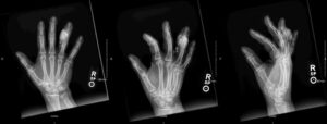
Figure 1. Preoperative X-ray of right hand demonstrating a large, calcified mass near the fourth digit middle phalanx and several smaller masses representing severe tumor calcinosis.
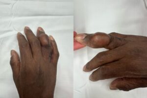
Figure 2. Preoperative picture of right hand showing the extent of tumor calcinosis finger deformity on ring finger.
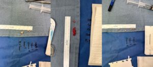
Figure 3. Multiple masses excised from right ring finger
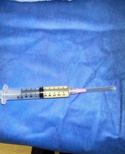
Figure 4. 5.5 mL of white, chalky fluid aspirated from the right ring finger.
Discussion
The term “Tumoral Calcinosis” has been used loosely over the years to describe any calcification of periarticular soft tissue. It was first used by Inclan, Leon, and Gomez (6) in 1943 to describe a dense, nodular, calcareous mass in the periarticular subcutaneous tissue. While the exact mechanism remains unclear, the cause in patients on hemodialysis is thought to be the result of abnormal metabolism of phosphate that forms high amounts of calcium-phosphorus products in the setting of secondary hyperparathyroidism (1).
The differential diagnoses for this patient included calciphylaxis, calcific tendonitis, and gouty arthritis. The absence of skin lesions, the presence of calcifications beyond the tendons, and normal uric acid levels, made these conditions less likely (7,8,9). Given the patient’s history of hemodialysis along with the identification of periarticular calcific lesions on x-ray and intraoperatively, the diagnosis of TC was made. In our review, all of the cases were met with surgical intervention, and no cases of recurrence were reported.
Our main objective in this case is to describe an uncommon presentation of a rare disease as well as demonstrate surgical treatment for a patient with TC of the hands. We would like to remind physicians to keep TC in their differential, even when it presents in uncommon locations. Future research should focus on identifying TC in other uncommon locations and determining a cause of this condition.
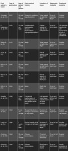
Table 1. Results of Pubmed search for similar cases of TC presenting in the hands
References
- Tumor Calcinosis – an overview | ScienceDirect Topics. Accessed April 2, 2024. https://www.sciencedirect.com/topics/pharmacology-toxicology-and-pharmaceutical-science/tumor-calcinosis
- Yano H. Tumoral calcinosis. doi:10.3949/ccjm.88a.20084
- Amati C, Pesce V, Armenio A, Solarino G, Moretti B. Tumoral calcinosis of the hand. Journal of Surgical Case Reports. 2015;2015(4). doi:10.1093/JSCR/RJV036
- Jakka S, Narayan R, Mishra M, Rana F, Laik JK. Idiopathic tumoral calcinosis – rare clinico pathological entity: A report of two cases. Journal of Clinical and Diagnostic Research. 2017;11(6). doi:10.7860/JCDR/2017/26779.10070
- Anderson KD, Malek F, Szewc RG, McCarthy WA, Hartzler RU. Tumoral calcinosis complicating a reverse total shoulder arthroplasty: a case report. JSES Reviews, Reports, and Techniques. 2022;2(1):87-91. doi:10.1016/j.xrrt.2021.09.005
- Inclan A, Leon P, Gomez Camejo M. Tumoral Calcinosis. JAMA. 1943;121(7):490-495. doi:10.1001/jama.1943.02840070018005
- Westphal SG, Plumb T. Calciphylaxis – StatPearls – NCBI Bookshelf. StatPearls Publishing. Published January 2024. Accessed April 2, 2024. https://www.ncbi.nlm.nih.gov/books/NBK519020/
- Kim MS, Kim IW, Lee S, Shin SJ. Diagnosis and treatment of calcific tendinitis of the shoulder. Clinics in Shoulder and Elbow. 2020;23(4):210. doi:10.5397/CISE.2020.00318
- Erickson TD, Assylbekova B, Chong ACM. Multiple Subcutaneous Gouty Tophi Even with Appropriate Medical Treatment: Case Report and Review of Literature. Kansas Journal of Medicine. 2021;14:12. doi:10.17161/KJM.VOL1414505
- Gonzalez M, Rettig M, Ayalon O. Extensive Tumoral Calcinosis of the Hand. Journal of Hand Surgery. 2021;46(11). doi:10.1016/j.jhsa.2020.10.030
- El Maghraoui J, Hammou M, Kabbali N, Arrayhani M, Houssaini TS. Amélioration de la calcinose tumorale de la main droite après para thyroïdectomie chez un hémodialysé chronique. Pan African Medical Journal. 2016;24. doi:10.11604/pamj.2016.24.30.8814
- Xu C, Potter JA, Carter CD, Lang CMC. Idiopathic tumoral calcinosis in hand: a case report. Eplasty. 2014;14.
- Kim HS, Suh JS, Kim YH, Park SH. Tumoral calcinosis of the hand: Three unusual cases with painful swelling of small joints. Archives of Pathology and Laboratory Medicine. 2006;130(4). doi:10.5858/2006-130-548-tcotht
- Amadei F, Petrolati M. Idiopathic tumoral calcinosis associated with congenital malformation of the hand. Case report. Annales de Chirurgie de la Main et du Membre Superieur. 1998;17(1). doi:10.1016/S0753-9053(98)80021-5
- Asuncion GF, Tzarnas CD. Uremic tumoral calcinosis: Acute hand presentations mimicking infection. Journal of Hand Surgery. 1994;19(5). doi:10.1016/0363-5023(94)90190-2
- Evans DM, Lewis JS. Tumoral calcinosis associated with congenital malformations of the hand. Journal of Hand Surgery. 1994;19(5). doi:10.1016/0266-7681(94)90136-8
