
Jonathan Tobin OMS-II; Shaunak Digambar OMS-II; Jessica Gilbertie PhD; Tiffany Carpenetti PhD
Edward Via College of Osteopathic Medicine, Virginia Campus
DOI: http://doi.org/10.70709/urnigcs
Abstract
This report highlights the presence of interlocking bilateral longitudinal grooves, resembling cogwheels, between the lateral femoral condyles and patellar articular surfaces in a 93-year-old female cadaver. To facilitate a thorough assessment of the articular surfaces and internal structures, bilateral knee joint dissections were initiated by sectioning the quadriceps tendons and reflecting the patellae inferiorly. Photographic documentation was used to illustrate these cogwheel grooves in the left and right patellofemoral joints. Remarkably, the cogwheel grooves were more pronounced in the right knee compared to the left, predominantly affecting the lateral femoral condyles and trochlear fossa of both knees. This report presents a unique and under-documented manifestation of degenerative joint disease. While the precise etiology of this degenerative joint pathology remains elusive, repeated knee flexion and extension, as suggested by the cogwheel track pattern, combined with compromised bone integrity, played a role in shaping this unique topographical presentation. Given the limited access to patient history, the influence of concurrent erosive arthropathies or autoimmune processes remains uncertain. Reporting this case offers valuable insights into an infrequently observed manifestation of degenerative joint disease. It assists healthcare professionals in gaining a deeper understanding of the disease’s progression, its severity, and indication for surgical intervention. To gain a more comprehensive understanding, future studies should expand by examining additional cadavers and conducting retrospective reviews of imaging studies. This should include the correlation of medical histories and demographic information with the occurrences of cogwheel grooves detected in the knee joint.
Keywords: Osteoarthritis, Degenerative Joint Disease, Knee, Patella
Introduction
Degenerative joint disease (DJD) due to osteoarthritis (OA) is the most common joint disease worldwide, impacting millions of individuals and posing significant challenges in clinical management. Degeneration begins when mechanical trauma inflicted upon the structures of a synovial joint elicits a biomechanical response in which the activation of cytokines builds an inflammatory response (1). These pro-inflammatory cytokines serve as mediators of matrix-degrading enzymes within the joint microenvironment. Consequently, a progressive erosion of cartilaginous structures ensues, accompanied by an escalating inflammatory response. Pathological findings in OA include several characteristic changes, including osteophyte formation, degeneration of cartilaginous tissues, alterations in subchondral bone architecture, synovial hyperplasia, and narrowing of the joint space (1). These hallmark manifestations collectively highlight the complex nature of OA pathogenesis, reflecting the intricate interplay between mechanical insults, inflammatory cascades, and subsequent tissue remodeling processes.
While classical presentations of OA are well-documented, atypical manifestations such as sawtooth grooving in the patellofemoral joint present unique diagnostic and therapeutic considerations. Previous reports have described this finding as being similar to overlapping cogs in a wheel or as having a sawtooth appearance (2,3). These bony changes arise from significant bone remodeling, resulting in distinctive grooves on the joint’s surface. The overall prevalence of sawtooth findings in the context of OA is unknown. The most frequently reported location of this degenerative feature is the knee joint (2,3). In the setting of erosive OA of the hand, sawtooth findings have been described in the proximal interphalangeal joints (4). While sawtooth OA has been previously described in the literature, it is overall infrequently reported. Recognizing and understanding an atypical presentation of OA such as sawtooth grooving is important for accurate diagnosis and effective treatment planning. This case report aims to present an unusual pattern of joint degeneration, characterized by sawtooth grooves observed bilaterally in the knee joints of a cadaver.
Case Presentation:
A 93-year-old female formalin-fixed cadaver was found to have interlocking bilateral longitudinal grooves located between degenerated lateral femoral condyles and patellar articular surfaces. Past medical history was unavailable; however, this individual was ambulatory until she suffered a hip fracture shortly before her death. Bilateral knee joint dissections were initiated through a longitudinal incision on the anterior surface of the knee joint. The sectioning of both quadricep tendons allowed for inferior reflection of patellae, thereby exposing the joint space. Full-thickness cartilage loss and bone eburnation were observed bilaterally in the knee joints. Sawtooth grooves present in the left and right patellofemoral joints were visually captured with photographic documentation. Subsequently, both the left and right distal femurs, patellae, and tibial plateaus were removed and placed in a Bruker SkyScan -1278 µCT scanner (100 μm) for additional analysis. While any clinical symptoms observed in the patient during their lifetime were unable to be attained, it is likely the patient would have experienced knee pain, stiffness, and/or functional limitations.
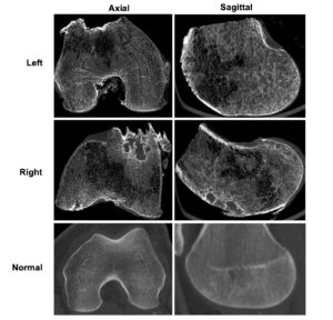
Image 1. µCT scans of right and left distal femurs, and CT scans of normal distal femur.
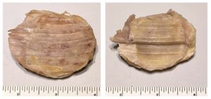
Image 2. Left and right dissected patella showing sawtooth degeneration on the articular surface.
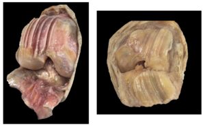
Image 3. Dissections of the right and left knee joints revealing sawtooth grooves in the patellofemoral joint space.
Discussion
There is a general lack of literature regarding the clinical implications of sawtooth OA cases, including management strategies and long-term prognosis. Previous studies have identified similar findings only in paleopathological specimens, likely due to the modern availability of surgical management of OA—an option that prevents excessive degeneration of knee joints (3,5). Evidence suggests that such cases have been found in patients with mild symptoms, those experiencing non-specific knee pain, and following a traumatic injury (2,3,5). Detecting OA early, with its many various presentations, is crucial for timely intervention and potentially enhancing patient outcomes. Based on the resemblance to previously documented cases, this donor likely experienced degenerative joint changes due to OA rather than an autoimmune origin or concurrent inflammatory arthropathies (6). However, definitive confirmation was not attainable due to the limited availability of detailed medical history. The apparent sawtooth pattern also suggests a potential for repetitive knee flexion and extension. Similar findings have been reported in female cadaveric specimens previously, suggesting that an increased valgus knee angle may contribute to its development (7). While sawtooth ridging in the knee joint has been documented in radiographs, the utilization of various imaging modalities, such as MRI and CT scanning, provides important insights into the relationship between clinical symptoms and structural abnormalities in DJD caused by OA (2). Imaging is useful for confirming a diagnosis of osteoarthritis and for evaluating the need for surgery.
Surgical management of OA is guided by current recommendations, which emphasize evidence-based approaches (8). Factors such as obesity, diabetes, and chronic pain can influence surgical outcomes, highlighting the need for personalized treatment decisions based on individual patient characteristics. Total knee arthroplasty (TKA) can be a viable option for patients with advanced OA, including those presenting with sawtooth grooving, with the goal of alleviating pain and restoring function. A recent systematic review and meta-analysis evaluated the clinical effectiveness and safety of TKA, examining various surgical outcomes and patient-reported measures (9). Among the seven different surgical approaches analyzed, the midvastus approach consistently yielded favorable outcomes both during and after surgery (9). Additionally, an examination of risk levels associated with TKA revealed a notable increase in procedures among patients under 65, with younger cohorts exhibiting higher failure rates primarily due to aseptic loosening (10). Age also emerged as a significant factor, with patients aged 50-65 showing a twofold increased risk of implant revision compared to older individuals. Alternative treatments, such as biological reconstruction and injective biological solutions, show promise in postponing or circumventing the need for TKA in younger patients with early to moderate OA (10). However, the demand for TKAs remains high across varying levels of OA severity, underscoring the importance of thorough evaluation and counseling regarding surgical indications.
Conclusion
In this study, we present the observation of bilateral grooving of the patellofemoral joints in a female cadaveric specimen. While sawtooth grooves are infrequently reported and typically associated with advanced stages of OA, this case underscores the significance of identifying atypical presentations of degenerative joint pathology. Furthermore, our study advances the potential use of µCT scans for exploring and evaluating OA manifestation in cadaveric specimens. By combining various imaging techniques with clinical data, we gain a deeper understanding of OA progression and potential contributing factors to the development of clinically relevant joint pathologies. Although the exact cause of this degenerative joint pathology remains unknown, surgical revision may have been indicated in this donor. Surgical management, particularly total knee arthroplasty (TKA), remains a viable option for patients with advanced OA. However, careful consideration of risks and benefits, particularly in younger patients, is essential. This highlights the importance of thorough evaluation and counseling regarding surgical interventions, ensuring individualized treatment plans tailored to each patient’s specific needs and circumstances.
References:
- Jang S, Lee K, Ju JH. Recent Updates of Diagnosis, Pathophysiology, and Treatment on Osteoarthritis of the Knee. Int J Mol Sci. 2021;22(5):2619. doi:10.3390/ijms22052619
- Anbarasu A, Loughran CF. Saw Tooth Patello — Femoral Arthritis. Clinical Radiology. 2000;55(10):767-769. doi:10.1053/crad.2000.0512
- Hardy E, Merrett DC, Zhang H, Zhang Q, Zhu H, Yang DY. Possible case of pressure resorption associated with osteoarthritis in human skeletal remains from ancient China. International Journal of Paleopathology. 2019;24:1-6. doi:10.1016/j.ijpp.2018.07.005
- Favero M, Belluzzi E, Ortolan A, et al. Erosive hand osteoarthritis: latest findings and outlook. Nat Rev Rheumatol. 2022;18(3):171-183. doi:10.1038/s41584-021-00747-3
- Rogers JM, Dieppe PA. Ridges and grooves on the bony surfaces of osteoarthritic joints. Osteoarthritis and Cartilage. 1993;1(3):167-170. doi:10.1016/S1063-4584(05)80087-1
- Paladichuk S, Gonzaga A, Lindsey J, Walser R. Cogwheel Grooves on Patellofemoral Articular Surfaces in a Cadaver. Pacific Northwest University of Health Sciences. Accessed March 2, 2024. https://www.pnwu.edu/2022-research-symposium-poster-hall/case-report-or-case-series/cogwheel-grooves-on-patellofemoral-articular-surfaces-in-a-cadaver/
- Emery IH, Meachim G. Surface morphology and topography of patello-femoral cartilage fibrillation in Liverpool necropsies. J Anat. 1973;116(Pt 1):103-120.
- McGrory BJ, Weber KL, Jevsevar DS, Sevarino K. Surgical Management of Osteoarthritis of the Knee: Evidence-based Guideline. J Am Acad Orthop Surg. 2016;24(8):e87-93. doi:10.5435/JAAOS-D-16-00159
- Zhao JL, Zeng LF, Pan JK, et al. Comparisons of the Efficacy and Safety of Total Knee Arthroplasty by Different Surgical Approaches: A Systematic Review and Network Meta-analysis. Orthop Surg. 2022;14(3):472-485. doi:10.1111/os.13207
- Perdisa F, Bordini B, Salerno M, Traina F, Zaffagnini S, Filardo G. Total Knee Arthroplasty (TKA): When Do the Risks of TKA Overcome the Benefits? Double Risk of Failure in Patients up to 65 Years Old. Cartilage. 2023;14(3):305-311. doi:10.1177/19476035231164733
- Hacking C. Normal knee CT: Radiology case. Radiopaedia. November 26, 2022. https://radiopaedia.org/cases/normal-knee-ct.
