
Jordan Read DO, ATC1; Benjamin Hammer DO1; Neil L. McNinch PhD(c.), MS, RN, PSTAT®2; Koan Heindel DO1
1Western Reserve Hospital
2McNinch Biostats, LLC
DOI: http://doi.org/10.70709/rmkeza6c0j
Abstract
Introduction
Superior patellar dislocation can be easily misdiagnosed, as it rarely occurs. Presentation includes a high riding patella, acute knee pain, inability to perform a straight leg raise, and a skin dimple deformity. We present the first statistical analysis of this patient population, as well as the 41st reported case of superior patella dislocation.
Methods
Data from our case was combined with data collected from a comprehensive literature review of published superior patellar dislocation cases. All analyses were conducted using STATA/BE 17.0, and results were interpreted at a Type I Error Rate of alpha = 0.05 level of statistical significance.
Results
The mean age was 55.4 (+/- 14.5) years old, with females representing over half of the sample (n=24, 58.5%). There was not a significant difference in age by sex or time period. The most commonly reported mechanism of injury was hyperextension (n=15, 36.6%) and direct trauma (n=15, 36.6%). Clinical exam finding included knee locked in extension (n=38, 92.7%), severe pain (n=28, 68.2%), and skin dimpling (n=14, 34.1%). The highest reported treatment group was closed reduction with sedation (n=11, 26.8%).
Discussion
There should be high clinic suspicion for superior patellar dislocation when a patient in their 5th or 6th decade of life presents with direct trauma or hyperextension of the knee, skin dimpling, and a knee locked in extension. Radiographs demonstrating the inferior pole of the patella perched on the femoral condyles, with classic clinic exam findings, are sufficient to make the diagnosis. First line treatment may include closed reduction, although spontaneous reduction may also occur. Early clinical diagnosis will lead to improved patient care, early intervention, and improved pain control.
Keywords: Superior Patellar Dislocation, Superior Dislocation of the Patella, Intra Articular Patella Dislocation, Patella Dislocation, Knee Pain
Introduction
Superior patellar dislocation is a rare diagnosis. It is commonly misdiagnosed as a patellar tendon rupture. (1,2) Superior patellar dislocations often occur in the 5th or 6th decades of life; however, there have been reports in young non arthritic knee joints. (3) Patients most commonly present following a trauma or hyperextension event with symptoms similar to patellar tendon rupture. History and exam findings include acute pain, inability to perform straight leg raise, high riding patella, and skin deformity.
The first reported case was presented by Watson-Jones in 1946. Discrepancy exists in the number of presented cases since that time. (4) Due to the rarity of superior patellar dislocation, early recognition and treatment may be difficult. Misdiagnosis may lead to delayed care and higher overall morbidity. We have found 40 reported cases of superior patellar dislocation. This is 12 more cases than sited by any previous article. (5) We present our case as the 41st reported superior patellar dislocation, and provide the first statical analysis of patient population and clinic presentation. The goal of this study was to provide greater detail regarding the demographic, physical exam findings, and treatment options of superior patellar dislocation for improved patient outcome.
Case presentation
A 65-year-old female presented after striking the anterior aspect of her right knee on a heavy metal beam while walking in her garage. She experienced immediate knee pain with the knee locked in extension, and inability to ambulate. She was taken to the emergency department for further evaluation. On physical exam, her knee was positioned in full extension. There was a visible deformity with a prominent superior patellar pole, and a sizable dimple just distal to the inferior pole of the patella. The patient was unable to perform a straight leg raise and felt severe pain with any attempt to flex the knee. The patient was neurovascularly intact.
Plain radiographs in the emergency department demonstrated a high-riding patella with the inferior pole perched on the anterior aspect of the distal femur (Figures 1 and 2). Subsequent MRI demonstrated an intact extensor mechanism with no evidence of patellar or quadriceps tendon injury (Figures 3, 4, and 5). The patient was discharged home with no attempt at manual reduction in the emergency department.
Follow up radiographs in office 2 days later demonstrated spontaneous interim reduction (Figures 6 and 7). Corticosteroid injection was provided for pain relief. Patient was instructed to maintain knee immobilizer while ambulating for two weeks and perform gentle range of motion several times per day. At the six weeks follow appointment patient was doing well with no residual pain or instability; no further intervention was required.
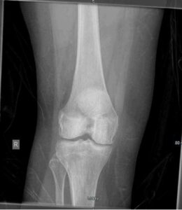
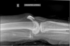
Figure 1 and 2. AP and lateral radiograph demonstrating superior patellar dislocation.
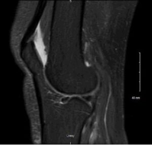
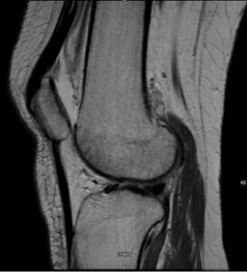
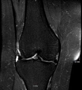
Figure 3, 4, and 5. Coronal and sagittal MRI demonstrating superior patellar dislocation with an intact patellar tendon.
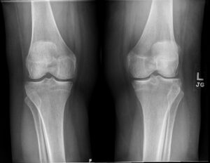
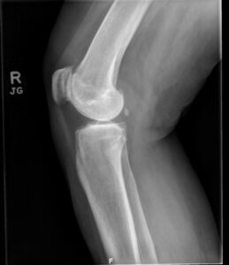
Figure 6 and 7. AP radiograph of bilateral knees and lateral radiograph of the right knee demonstrating reduced superior patellar dislocation.
Methods
Inclusion and exclusion criteria
Literature review was performed by librarian and key authors utilizing Google Scholar, Pubmed, EMBASE, and sited sources from previous superior patellar dislocation case reports. A key word search included the terms “superior patellar dislocation”, “superior patella dislocation”, “superior dislocation of the patella”, and “intra-articular patella dislocation”. Cited sources of each article found with a superior patellar dislocation case were reviewed. Case reports were gathered with inclusion of any subject that has sustained superior patellar dislocation.
Statistical Analysis
Data was collected from the current case and combined with data obtained through literature review. Analysis of data comprised of distribution-based summary statistics, overall and stratified by sex and year (pre/post 2000) [mean (standard deviation) for normally distributed covariates; n (%) for categorical data]. The Independent Samples T-Test was used to assess potential differences in age by sex and year (pre/post 2000). The Chi-Square Test of Independence (or Fisher’s Exact Test in cases of small cell counts, n < 5) was used to assess potential associations between categorical demographic and clinical characteristics with sex and year (pre/post 2000). All analyses were conducted using STATA/BE 17.0, and results were interpreted at a Type I Error Rate of alpha = 0.05 level of statistical significance.
Results
We analyzed and reported on 41 cases of superior patellar dislocation were identified. (1-36) There were 13 cases prior to the year 2000 (31.7%), and 28 cases were after 2000 (68.3%). The mean (sd) age was 55.4 (14.5) years old, with females representing over half of the sample (n=24, 58.5%). There was not a significant difference in age by sex (p-value = 0.683) or time period (p-value = 0.127), however the mean (sd) age prior to 2000 was 59.6 (9.3) years versus 53.4 (16.1) post 2000. There have been two reports in patients as young as 19 years old. (3,6) There was no evidence of association between sex and time epoch (p-value = 0.678). (Table 1)
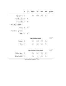
Table 1. Summary statistics and results of tests for difference of demographic characteristics.
Clinical characteristics examined included patient presentation, mechanism of injury, and treatment received. There were 15 patients (36.6%) reported to have sustained a superior patellar dislocation with hyperextension and 15 (36.6%) with direct trauma. (1,3-29) There were 5 reports (12.2%) of individuals who sustained this injury with a combination of hyperextension and direct trauma. (2,30-32) Sudden onset was reported for the remaining 6 cases (14.6%). (31,33) We found 11 reported cases (26.8%) of closed reduction with sedation as the most sighted treatment. (2-4,6,8,11-14,21,23,28,31,33) There were 10 cases (24.4%) of closed reduction without anesthesia. (1,7-9,18,19,31) Only one case (2.4%) presented required open reduction.30 (Table 2)
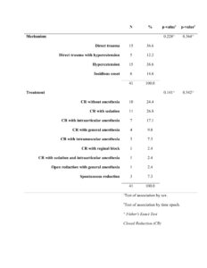
Table 2. Frequency distributions and results for tests of association for clinical characteristics and treatment.
The most frequent clinical exam finding was a knee locked in extension, reported in 38 cases. (4,7-12,14,19,23,30,31) The next most reported clinical exam finding was severe pain, reported in 28 cases. (1-3,5,7-11,13,14,16,17,21,23-25,28-34) Skin dimpling just inferior to the superior dislocated patella was reported by 14 cases. (1,2,8,12,13,16-18,21,27,28,32,34) Only 12 studies reported testing for active straight leg raise and patient ambulatory status, 8 (66.7%) of which were negative for ability to perform straight leg raise or ambulate. (7,9,10,12,14,30) There were no significant associations for ability to straight raise leg (p-value = 0.208) or ambulate (p-value = 0.081) by sex. (Table 3)
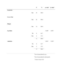
Table 3. Frequency distributions and results of tests for association of presenting symptoms by sex and time epoch.
Discussion
Our literature review found 40 previously reported cases of superior patellar dislocation. (1-36) As superior patella dislocation is a rare diagnosis, accurate reporting aids in providing more precise information for clinical diagnosis and outcomes. It has been postulated the number of superior patellar dislocation cases would increase as the population age increases and more people develop osteoarthritis. (2,13,21) We found 28 cases reported after the year 2000 compared to 13 reported prior to the year 2000. This may be evidence that superior patellar dislocation is becoming more widely recognized and reported. We found the average age to be 55.4 years old, with no statical difference between sex. This is consistent with prior literature, which states that superior patellar dislocation commonly occurs in the 6th decade of life. (5,7,9,11,13,17,32) The mean (sd) age prior to 2000 was 59.6 (9.3) years versus 53.4 (16.1) post 2000, this decrease in reported age was not found to be statically different but it may be due to the fact of several recently reported cases occurring with young individuals without osteoarthritis or osteophyte formation.
A knee locked in extension was the most sighted physical exam finding for superior patellar dislocation, and is important to distinguish other pathology of the knee. (4,7-12,14,19,23,30,31) Locking often occurs as the inferior poll of the patella interlocks on the superior ridge of the infrapatellar groove. This has been reported as osteophyte formation increases the surface area of the inferior pole of the patella and the femoral ridge. This locking mechanism may be the cause of severe pain and other clinical exam findings.
Severe pain was the second most reported clinical exam finding. (1-3,5,7-11,13,14,16,17,21,23-25,28-34) Pain is likely caused by the inappropriate bone-on-bone articulation; as well as acute inflammation and ligament disfunction. As severe pain is not specific to superior patella dislocation it does not differentiate from other knee pathology. It does indicate that these patients have acute and significant pain and are likely to present to an emergency room, urgent care, or primary care clinic. This stresses the importance of multiple disciplinary recognition and diagnosis of this unique pathology.
The skin dimpling pattern of superior patella dislocation is a unique exam finding specific for superior patella dislocation. (1,2,8,12,13,16-18,21,27,28,32,34) A skin dimple is formed when the superiorly dislocated patella inverts with the distal portion angled posteriorly applying tension to the intact patellar tendon and skin, which dimples distally. Van Egmond et al. presented that this skin dimple sign may be related to the radiographic finding of the superior poll of the patella tilting away from the femur as the intact patellar tendon is being stretched. They described this radiographic patellar tilt as being pathognomonic for a superior patellar dislocation. (16) If further evaluation is required, ultrasound or MRI may be performed to further evaluate the integrity of the patellar tendon and other associated pathology.
Inability to perform straight leg raise or ambulate are exam findings coincident with patellar tendon rupture, making it difficult to differentiate between the two diagnoses. Few studies reported inability to perform straight leg raise and inability to ambulate. (7,9,10,12,14,30) Inability to straight leg raise or ambulate is likely secondary to severe pain. It is possible for quadriceps function to be inhibited by the interlocking patella mechanics or stretching the patellar tendon. As a clinician it is important to perform a thorough physical exam and differentiate the cause of a patient’s inability to perform diagnostic tests and rule out other associated pathology.
We found the most common mechanisms of injury were direct trauma and hyperextension. (1,3-29) This is consistent with prior literature. Takai et al. described a patient who was able to voluntarily perform superior patellar dislocation with hyperextension from a slightly flexed position. (19) A thorough history is vital to accurate diagnosis and differentiate the diagnosis of superior patella dislocation when a patient reports a knee injury caused by direct trauma or hypertension. {Takai, 1998 #156}
In our case spontaneous reduction without manipulation of the dislocation occurred. Many reported treatment options for superior patella dislocation exists. We found three reports of spontaneous reduction without intervention. (16,31) The most commonly reported treatment was closed reduction with sedation. (2-4,6,8,11-14,21,23,28,31,33) Ten cases of closed reduction were performed without anesthesia. (1,7-9,18,19,31) Seven cases were treated with intraarticular anesthesia. (1,10,15,17,25,27,32) Yip et al. presented an algorithm for the treatment of superior patellar dislocation which instructs clinicians to begin by performing closed reduction with simple anesthesia. (31) Based on our findings treatment should be clinician and practice dependent and may include sedation or intraarticular anesthesia as needed. Other options included intramuscular anesthesia or general anesthesia. If closed reduction is unsuccessful or recurrence occurs, patients have been successfully treated with an arthroscopic or open procedures. (19,30)
Limitation to this analysis included the small sample size of convenience, therefore results of this analysis may not have been powered at the typically desired level of 80%, meaning power was too low to detect effects should they be present. Additional limitations lie in the use of pooled individual data from literature. The results of this analysis are not necessarily generalizable but may serve to inform future research.
Conclusion
We present the 41st reported case of superior patella dislocation. It is a rare diagnosis that typically occurs in the sixth decade of life. It can be easily misdiagnosed as a patellar tendon rupture secondary to severe pain, high riding patella, in ability to ambulate or perform a straight leg raise. Our statistical analysis did not demonstrate any statistically significant results; however, it did support previous literature which reported injury often occurs in the 6th decade of life with direct trauma or hyperextension mechanism of injury. Any clinical provider who has a patient that presents with their knee locked in extension and skin dimpling should have a high suspicion for superior patella dislocation. Knee locked in extension was the most sited clinical exam finding and is highly suggestive of superior patella dislocation. Radiographs demonstrate the inferior pole of the patella perched on the femoral condyle and are sufficient to confirm the diagnosis. Closed reduction has been a successful first line treatment with surgery as an option for patients with recurrence or bone fragments.
References
- Boonrod A, Sumanont S, Boonard M, Boonrod A. Superior patellar dislocation misdiagnosed as patellar tendon rupture: the value of ultrasonography. Case reports in orthopedics. 2016;2016.
- Bassi R, Kumar B. Superior dislocation of the patella; a case report and review of the literature. Emergency Medicine Journal. 2003;20(1):97-98.
- Kataoka T, Iizawa N, Takai S. Superior dislocation of the patella in a young woman without osteophytes: a case report. Journal of Nippon Medical School. 2016;83(1):24-26.
- Watson-Jones R. Fractures and Joint Injuries. Vol 2. 3rd ed. Baltimore, Md, USA: Williams and Walkins; 1946.
- Berry CA, Summers MA, Reddy KI. Superior Patellar Dislocation Reduced by Intra-articular Injection: A Case Report and Review of Literature. JBJS Case Connector. 2021;11(2):e20.
- Brito WEd, Campos GC, Miranda JBd, Zorzi AR. Superior Dislocation of the Patella in a Young Patient without Osteophytes: A Case Report with Discussion about Differential Diagnosis. Case Reports in Medicine. 2019;2019:7314698.
- Bartlett D, Gilula L, Murphy W. Superior dislocation of the patella fixed by interlocked osteophytes. A case report and review of the literature. JBJS. 1976;58(6):883-884.
- Wimsatt MH, Edward JCJ. Superior Dislocation of the Patella. Journal of Trauma and Acute Care Surgery. 1977;17(1):77-79.
- Siegel MG, Mac SS. Superior dislocation of the patella with interlocking osteophytes. The Journal of trauma. 1982;22(3):253-254.
- Hanspal R. Superior dislocation of the patella. Injury. 1985;16(7):487-488.
- Friden T. A case of superior dislocation of the patella. Acta Orthopaedica Scandinavica. 1987;58(4):429-430.
- Teuscher DD, Goletz TH. Recurrent atraumatic superior dislocation of the patella: case report and review of the literature. Arthroscopy: The Journal of Arthroscopic & Related Surgery. 1992;8(4):541-543.
- Scott S, Molloy A, Harvey R. Superior dislocation of the patella—a rare but important differential diagnosis of acute knee pain—a case report and review of the literature. Injury. 2000;31(7):543-545.
- McWilliams T, Binns M. A locked knee in extension: a complication of a degenerate knee with patella alta. The Journal of bone and joint surgery British volume. 2000;82(6):890-890.
- Ofluoglu O, Yasmin D, Donthineni R, Yildiz M. Superior dislocation of the patella with early onset patellofemoral arthritis: a case report and literature review. Knee Surgery, Sports Traumatology, Arthroscopy. 2006;14(4):350-355.
- van Egmond P, Vermeulen M, van Dijke C, Graat H. Superior dislocation of the patella: a pathognomonic finding and review of literature. Skeletal radiology. 2017;46(2):259-264.
- Joseph G, Devalia K, Kantam K, Shaath NM. Superior dislocation of the patella. Case report and review of literature. Acta Orthop Belg. 2005;71(3):369-371.
- Saleemi AJ, Hussain A, Iqbal MJ, Thuse MG, George AA. Superior dislocation of patella in a rugby player: an update on a extremely rare condition and review of literature. Knee Surgery, Sports Traumatology, Arthroscopy. 2007;15(9):1112-1113.
- Takai S, Yoshino N, Hirasawa Y. Case report arthroscopic treatment of voluntary superior dislocation of the patella. Arthroscopy: The Journal of Arthroscopic & Related Surgery. 1998;14(7):753-756.
- Iorwerth A, Thomas R, Shewring D. Confirmation of an intact patellar tendon in superior dislocation of the patella using magnetic resonance imaging. Injury. 2001;32(2):167-169.
- Clift RK, El-Alami W. Superior patellar dislocation: the value of clinical examination and radiological investigation. Case Reports. 2012;2012:bcr2012007571.
- Garner JP, Pike JM, George CD. Intra-articular dislocation of the patella: two cases and literature review. Journal of Trauma and Acute Care Surgery. 1999;47(4):780.
- Roth RM, McCabe JB. Nontraumatic superior dislocation of the patella. The Journal of Emergency Medicine. 1985;3(4):265-267.
- Lai R, Lau Y. Superior dislocation of the patella treated by closed reduction: a rare case report and review of the literature. Hong Kong Journal of Emergency Medicine. 2007;14(4):225-227.
- Cusco X, Seijas R, Ares O, Cugat JR, Garcia-Balletbo M, Cugat R. Superior dislocation of the patella: a case report. Journal of Orthopaedic Surgery and Research. 2009;4(1):29.
- Hansen B, Beck C, Townsley R. Arthroscopic removal of a loose body osteophyte fragment after superior patellar dislocation with locked osteophytes. Arthroscopy: The Journal of Arthroscopic & Related Surgery. 2003;19(3):1-4.
- YOSHINO N, TAKAI S, NAKAMURA S, MANABE T, HIRASAWA Y. Recurrent Horizontal Dislocation of the Patella in the Sagittal Plane. A Case Report*. JBJS. 1996;78(2):278-280.
- Glasbrenner J, Briese T, Raschke MJ, Herbst E, Kittl C. [A rare cause of an acutely locked knee joint: superior patellar dislocation]. Unfallchirurg. 2021;124(5):407-411.
- Siddiqui MA, Tan M. Locked knee from superior dislocation of the patella-diagnosis and management of a rare injury. Knee Surgery, Sports Traumatology, Arthroscopy. 2011;19(4):671-673.
- Rao J, Meese M. Irreducible superior dislocation of the patella requiring open reduction. American journal of orthopedics (Belle Mead, NJ). 1997;26(7):486-488.
- Yip D, Wong J, Sun L, Wong N, Chan C, Lau P. The management of superior dislocation of the patella with interlocking osteophytes—an update on a rare problem. Journal of orthopaedic surgery. 2004;12(2):253-257.
- Brown MJ, Ahn R, Nenno DJ. Locked Superior Patellar Dislocation. Orthopedics. 2016;39(2):e359-e361.
- Jahangir N, Umar M. Spontaneous superior patellar dislocation in young age: case report and reduction technique. Journal of surgical case reports. 2017;2017(3):rjx036.
- Dubey V, Sangnod PA, Samant A, Shahane SM. Superior Dislocation of Patella – A RareClinicalEntity. J Orthop Case Rep. 2019;9(2):42-44.
- Gakhar H, Singhal A. Superior dislocation of the patella: case report and review of the literature. Journal of Emergency Medicine. 2013;44(2):478-480.
- Javedani PP, Goldberg LC, Panchal AR. Closed Emergency Department Reduction of a Superior Patellar Dislocation After Blunt Trauma. The Journal of Emergency Medicine. 2018;55(4):567-569.
