
Michael O’Dell, OMS 20271; Arash Badragheh, OMS 20271; Jason Fink, OMS 20271; Stephen Cairone, DO2
1Philadelphia College of Osteopathic Medicine, Philadelphia, PA
2Trinity Health Mid-Atlantic Medical Group, Langhorne, PA
These authors contributed equally to this study and should be considered equal first authors.
Abstract
Background: Multiple myeloma frequently involves the spine, with approximately 47% of patients developing vertebral compression fractures. Although most spinal involvement occurs in the thoracolumbar region, lesions affecting the upper cervical spine pose unique challenges because the atlantoaxial articulation plays a critical role in maintaining craniocervical stability. Cervical spine instability from multiple myeloma is infrequently reported, and surgical management of atlantoaxial involvement remains underrepresented in the literature.
Case presentation
A 63-year-old male with multiple myeloma presented to the emergency department with syncope, dizziness, and cervicalgia, found to have a pathologic C2 vertebral body fracture with a 3.0cm×2.1cm×1.5cm lytic lesion causing anterior displacement of the left lateral mass and concern for atlantoaxial instability. Despite completing radiation therapy and receiving chemotherapy with bortezomib and denosumab, no symptomatic improvement was observed. The Spinal Instability Neoplastic Score was 18, indicating definite instability confirmed by imaging and requiring surgical intervention. A posterior occiput to C5 fusion was performed using an occipital plate with bi-cortical screw fixation, bilateral C1 lateral mass screws, and lateral mass screws at C3–C5 with bilateral pre-contoured rods. Intraoperative imaging confirmed appropriate hardware placement. The patient experienced meaningful symptomatic improvement following surgery, with successful stabilization of the craniocervical junction.
Conclusion
This case demonstrates the effectiveness of posterior occipitocervical fusion for managing atlantoaxial instability secondary to multiple myeloma when conservative treatments fail. Clinicians should consider early surgical stabilization in patients with multiple myeloma affecting the C1–C2 articulation who demonstrate imaging evidence of instability and inadequate response to nonoperative management.
Keywords: Multiple myeloma, cervical spine, atlantoaxial instability, posterior cervical fusion, case report
Introduction
Multiple myeloma is a form of cancer that arises from the malignant transformation of plasma cells, leading to increased osteoclastic activity, lytic bone lesions, and pathologic fractures. (1,2) The increased osteoclast activity and lytic bone lesions may lead to weakening of the bone and ultimately result in fractures. Lytic lesions in the C2 vertebra may result in atlanto-axial subluxation, risking complications such as spinal cord compression and vertebral artery dissection. (3) Surgical interventions to address instability include occipitocervical fusion, posterior cervical decompression, and posterior cervical fusion.
Case Report
A 63-year-old male presented to the emergency department with multiple episodes of syncope, dizziness, and chronic neck pain that had acutely worsened following a syncopal fall. No relevant family or psychosocial history was identified. Imaging obtained during this workup revealed a closed nondisplaced pathologic fracture of C2 with an anterior epidural tumor extension, prompting further evaluation. A diagnosis of multiple myeloma was confirmed the following day based on CTA head/neck findings demonstrating a pathologic C2 fracture with an expansile rib lesion, in conjunction with an elevated total protein of 9.8 g/dL. At the time of diagnosis, the patient’s disease was classified as stage III per the Revised International Staging System (R-ISS), based on elevated total protein and the presence of multiple lytic bone lesions. The patient was subsequently seen in clinic, at which time he had been started on radiation therapy for the C2 lesion, denosumab 120mg, and weekly bortezomib 3.5mg, with a Miami J cervical collar for stabilization. Clinically, his cervical motion was unrestricted in all planes, and both upper extremities were normo-reflexic and without motor deficit. X-rays, CT, and MRI of the cervical spine revealed degenerative changes with severe stenosis at C6-C7 and a 3.0cm× 2.1cm×1.5cm lytic lesion in the body of C2 with pathologic fracture and anterior displacement of the left lateral mass, causing concern for instability (Figure 1-3). The clinical presentation of progressive cervicalgia, dizziness, and syncope in the setting of known multiple myeloma directed the diagnostic assessment toward a pathologic fracture with atlantoaxial instability. The characteristic lytic morphology on imaging, pathologic fracture pattern, and elevated total protein (9.8 g/dL) consistent with his established diagnosis supported myelomatous destruction of C2 as the primary etiology. Differential diagnoses considered included traumatic fracture from the syncopal fall, metastatic disease from an occult solid tumor primary, and vertebral osteomyelitis; these were excluded based on the absence of a primary solid tumor, lack of infectious signs, and imaging characteristics inconsistent with benign or traumatic etiologies. The Spinal Instability Neoplastic score was calculated at 18, indicating definite instability and necessitating surgical intervention, as the patient had already completed a full course of radiation therapy. A stage 1 posterior occiput-to-C5 fusion was performed to control the extensive atlantoaxial instability, with plans for a later stage 2 anterior C5-C7 decompression and fusion if still symptomatic due to degenerative changes at these levels.
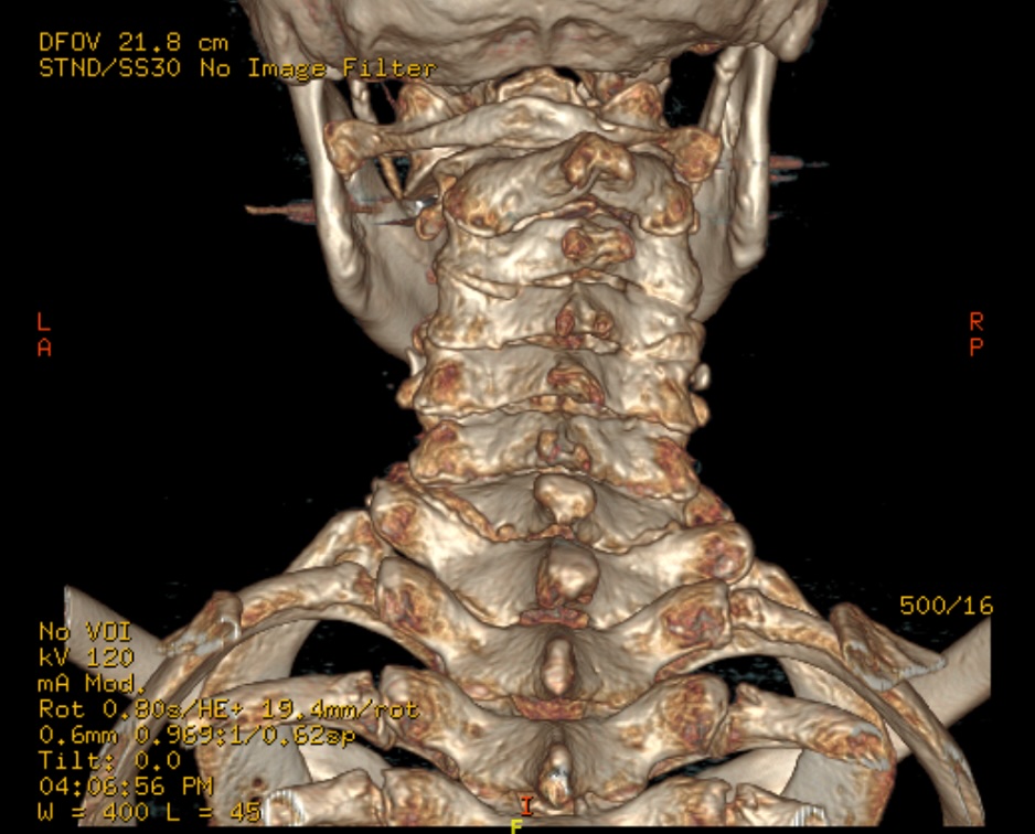
Figure 1: 3D coronal CT of a 63-year-old male with multiple myeloma demonstrating anterior displacement of the left C1-C2 lateral mass and atlantoaxial malalignment secondary to lytic destruction of C2.

Figure 2: Coronal CT demonstrating a large lytic lesion at the C2 vertebral body with C1-C2 instability secondary to multiple myeloma.
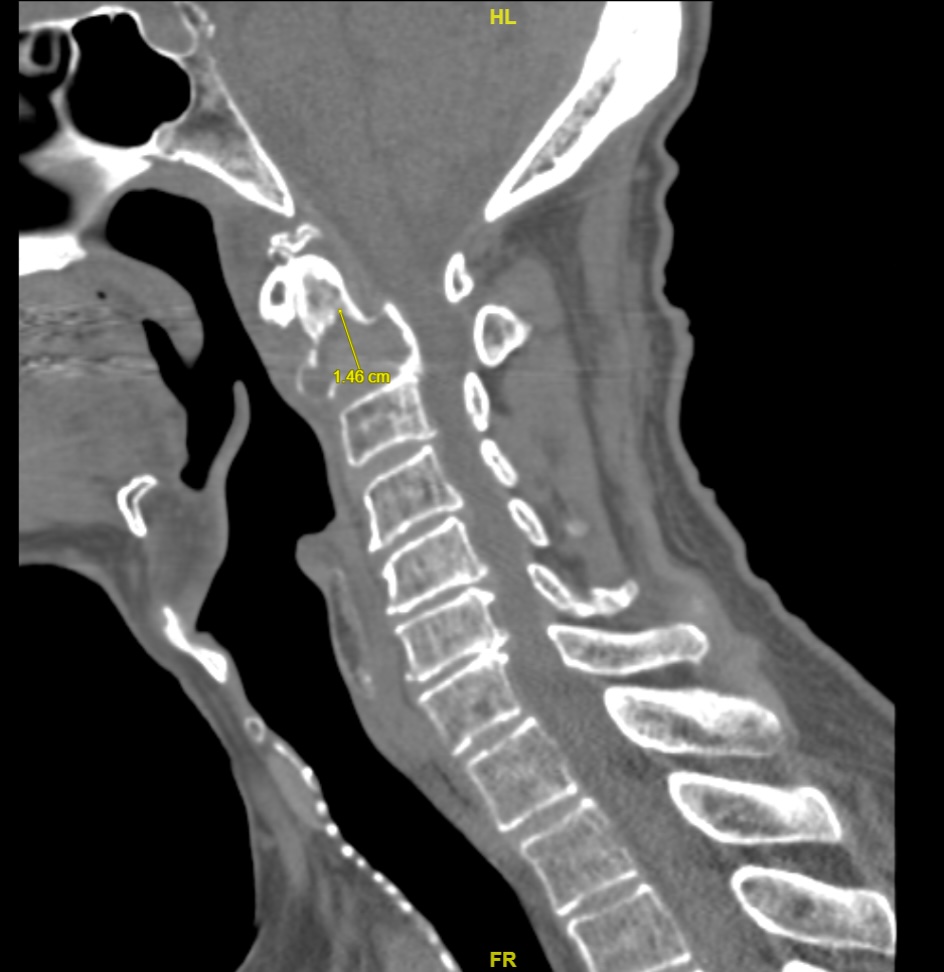
Figure 3: Sagittal CT demonstrating a lytic lesion of the C2 vertebral body measuring 3.0×2.1×1.5cm secondary to multiple myeloma.
An awake fiberoptic intubation was carried out to maintain control of the patient’s unstable cervical spine. A Mayfield head holder was used to maintain the neck in a neutral position during the fusion, and neuromonitoring leads were used to monitor the extremities.
A posterior midline incision was made, and the vertebral arteries were then identified and protected. A subperiosteal dissection was done from C1 to C5. The inion was skeletonized. An occipital plate was secured with bi-cortical screw fixation. A ball-tip probe was used to confirm dural integrity after each screw insertion. Bilateral C1 lateral mass bi-cortical screws were placed under fluoroscopic guidance. The right C2 nerve root was sacrificed to facilitate access for C1 lateral mass screw placement, after which a 45mm screw was inserted at the correct starting point and confirmed on lateral fluoroscopic imaging. Lateral mass screws were added at C3, C4, and C5, and bilateral pre-contoured rods were secured from the occiput to C5 using a rod reduction technique. The lateral masses were thoroughly decorticated, and the fusion bed was augmented with demineralized bone matrix and autograft to include the occipital bone. At the completion of the surgery, AP and lateral X-rays confirmed proper positioning of all hardware. A large Hemovac drain was placed subfascially, and the wound was subsequently closed.
Intraoperative AP and lateral X-rays confirmed appropriate hardware placement (Figure 4-5). Following 2 L of saline irrigation, a subfascial Hemovac was placed. Three weeks postoperatively, the patient was doing excellently and only reported minimal numbness and tingling in the right side of the posterior aspect of the head due to the need to sacrifice the C2 nerve root on the right side during the surgery. On X-ray, the hardware appeared well-placed with no signs of displacement. The patient was placed in a soft collar and tolerated it well. By six weeks postoperative, the patient continued to show improvement and was able to discontinue the soft collar. Throughout his care, he experienced no diagnostic challenges, barriers, or difficulties accessing care. He was fully compliant and tolerated his diagnosis and management well. Prior to surgery, there was consideration for the need to perform a follow-up surgery of a C5-C7 anterior decompression and fusion to address degenerative changes at these levels; however, the primary surgical objective was stabilization of the C2 pathology, with anterior intervention reserved for persistent symptoms attributable to the lower cervical disease. Because the patient improved and had no symptoms, it was determined that another surgery would provide no benefit. This surgery successfully stabilized the unstable articulation of C1-C2, enabling the patient to live without needing to wear a cervical collar for the remainder of his life. No future adverse events were anticipated at this point in the postoperative period. Table 1 summarizes the timeline of this patient’s care.
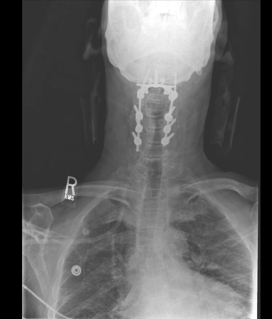
Figure 4: Intraoperative AP X-ray confirming posterior occiput-to-C5 fusion with occipital plate, bilateral C1 lateral mass screws, lateral mass screws at C3–C5, and bilateral pre-contoured rods.
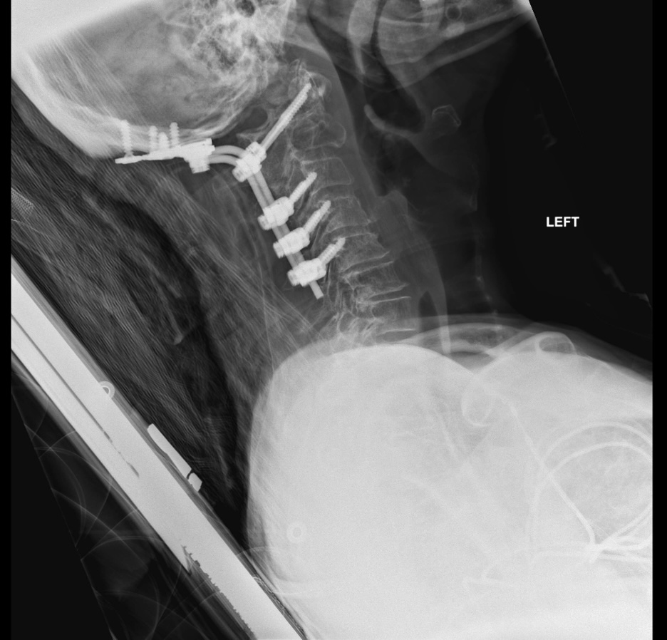
Figure 5: Intraoperative lateral X-ray confirming appropriate hardware placement following posterior occiput-to-C5 fusion for atlantoaxial instability secondary to multiple myeloma.
| Timepoint | Event |
| Initial ED presentation | 63-year-old male presenting to the ED with multiple episodes of syncope, dizziness, and chronic neck pain acutely worsened after a syncopal fall; cervical motion unrestricted; upper extremities normoreflexic and without motor deficit; imaging (X-ray, CT, MRI) demonstrating 3.0×2.1×1.5cm lytic lesion at C2 with pathologic fracture and anterior displacement of left lateral mass; SINS = 18 (definite instability) |
| Prior to presentation to our clinic | Diagnosis of multiple myeloma; 10 sessions of targeted radiation to C2 lesion; initiation of weekly bortezomib 3.5mg and single-dose denosumab 120mg; fitted with Miami JTO cervical collar |
| Surgical intervention | Posterior occiput-to-C5 fusion: awake fiberoptic intubation, occipital plate with bicortical screw fixation, bilateral C1 lateral mass screws, lateral mass screws C3–C5, bilateral pre-contoured rods; intraoperative imaging confirmed hardware placement |
| 3 weeks postoperative | Patient doing excellent; minimal numbness and tingling over right posterior scalp (expected, secondary to right C2 nerve root sacrifice); hardware well-positioned on X-ray; transitioned to soft cervical collar |
| 6 weeks postoperative | Continued improvement; soft collar discontinued; patient ambulatory without restriction |
Table 1: Timeline summary of the patient’s care.
Discussion
Vertebral multiple myeloma lesions are at risk of causing significant spinal instability. This may occur because destructive lytic activity weakens the spine’s structural integrity, predisposing affected levels to pathologic compression fractures and subsequent collapse. (1,4) Vertebral involvement in multiple myeloma is prevalent, with approximately 47% of patients developing compression fractures. (3,5)
When these lesions involve the upper cervical spine, particularly C1 and C2, the risk increases because the atlantoaxial complex plays a critical role in maintaining craniocervical stability. In this case, extensive lytic destruction of the C2 vertebral body resulted in a pathologic compression fracture with associated retropulsion of fragments and anterior translation of C1 on C2, leading to marked narrowing at the craniocervical junction and mechanical instability. Additionally, extension of the myelomatous lesion into the epidural space, along with pressure from the collapsed C2 vertebra, likely contributed to spinal cord deformation at the cervicomedullary junction, further compromising neural function. These structural changes were likely a significant contributor to the initial onset and flare-ups of the patient’s symptoms, which included worsening neck pain, dizziness, and recurrent syncopal episodes.
Although nonoperative management may be effective for certain patients, surgical intervention remains indicated in the presence of cervical instability or structural collapse, particularly when conservative measures have failed. (6,7) In this case’s patient, no therapeutic response or symptomatic improvement was observed following 10 sessions of targeted radiation to the C2 lesion, along with no measurable benefit from concurrent chemotherapy.
The Spinal Instability Neoplastic Score (SINS) is a validated classification system assessing tumor-related spinal instability across six components, with scores of 13–18 indicating definite instability warranting surgical consultation. (8) In this patient, a SINS of 18 reflected a junctional lytic lesion at the occiput–C2 region with subluxation, vertebral body collapse, and posterolateral involvement, confirming definite instability following failure of conservative management.
The surgical approach for treating multiple myeloma involving the atlantoaxial region parallels that for metastatic tumors, as both present with similar mechanical instability and neurological complications. (9) Two-staged anterior retropharyngeal excision with posterior stabilization achieves direct decompression but carries significantly higher perioperative morbidity, making it poorly tolerated in patients with compromised medical status. (10,11)
Vertebral augmentation via vertebroplasty and kyphoplasty is not routinely performed at C1–C2 because of the small vertebral bodies, proximity to critical neurovascular structures, and reported cement extravasation rates up to 60%. (12,13,14) Furthermore, while vertebral augmentation provides effective pain relief in 75-100% of multiple myeloma patients at thoracolumbar levels, it does not address mechanical instability or provide adequate decompression when cord compression is present. (15)
In this patient, because conservative treatments did not provide stability or symptom relief and imaging showed clear instability at the atlantoaxial joint, an occipitocervical fusion was considered necessary for definitive stabilization. This broader construct provided the rigidity needed to control craniocervical motion and prevent further displacement, an outcome that isolated C1–C2 decompression or more limited segmental fixation would not have achieved. During the procedure, the right C2 nerve root was sacrificed to allow unobstructed access for C1 lateral mass screw placement, resulting in a manageable area of occipital sensory deficit without significant functional consequences. This approach successfully stabilized the craniocervical junction, relieved the ongoing pressure on the spinal cord, and led to clear, meaningful improvement in the patient’s symptoms.
A limitation of this study is the lack of postoperative standing radiographs for cervical alignment assessment, as imaging was obtained at an outside facility and records were not available for inclusion at the time of manuscript preparation. Additionally, as a single-patient case report, this study is inherently limited in sample size. While the patient tolerated the management well and achieved sufficient cervical stability to discontinue his soft collar, the generalizability of this approach to cervical spine instability secondary to multiple myeloma remains limited, and further studies with larger patient cohorts are needed to validate these findings.
Conclusion
The risks of atlantoaxial subluxation include nerve compression, paralysis, and even death. Surgical correction is indicated for chronic or severe instability, reducing the risk of severe complications. While multiple myeloma is not a common cause of subluxation, this case shows that when the disease affects the C1-C2 joint, there is a risk of instability and possible subsequent subluxation. (8) Posterior cervical fusion and occipitoatlantal fusion may be indicated surgical procedures for stabilizing the cervical spine. Cervical instability may be caused by a wide variety of traumatic injuries, chronic disorders, and diseases. Multiple myeloma as the cause of cervical spine instability is infrequently reported compared to other etiologies. This case illustrates the advantages of surgical fixation for atlantoaxial instability in patients with multiple myeloma affecting the C1-C2 articulation.
Funding
No funding was received for this work.
Conflicts of Interest
The authors declare no competing interests.
Availability of Data and Materials
Not applicable.
Authors’ Contributions
Conceptualization: MO and JF; methodology: MO and JF; validation: MO, JF and AB; formal analysis: MO, JF and AB; investigation: MO, JF and AB; writing-original draft preparation: MO, JF and AB; writing-review and editing: all authors; visualization: MO and JF; supervision: SC. All authors have read and agreed to the published version of the manuscript.
Ethics Approval Statement
Not applicable.
Consent for Publication
Informed consent was obtained for publication of this case report and accompanying images.
References
- Esteve FR, Roodman GD. Pathophysiology of myeloma bone disease. Best Pract Res Clin Haematol. 2007;20(4):613–24. doi:10.1016/j.beha.2007.08.003
- Abduh MS. An overview of multiple myeloma: a monoclonal plasma cell malignancy’s diagnosis, management, and treatment modalities. Saudi J Biol Sci. 2024;31(2):103920. doi:10.1016/j.sjbs.2023.103920
- Zijlstra H, Wolterbeek N, Ponds NHM, Koene HR, Terpstra WE, Delawi D, et al. The incidence of vertebral compression fractures and spinal instability in newly diagnosed multiple myeloma patients. J Orthop. 2023;38:62–7. doi:10.1016/j.jor.2023.03.008
- Quidet M, Zairi F, Boyle E, Facon T, Vieillard MH, Machuron F, et al. Evaluation of the relevance of surgery in patients with multiple myeloma harboring symptomatic spinal involvement: a retrospective case series. World Neurosurg. 2018;114:e356–65. doi:10.1016/j.wneu.2018.02.184
- Lecouvet FE, Vande Berg BC, Maldague BE, Michaux L, Laterre E, Michaux JL, et al. Vertebral compression fractures in multiple myeloma. Part I. Distribution and appearance at MR imaging. Radiology. 1997;204(1):195–9. doi:10.1148/radiology.204.1.9205246
- Sciubba DM, Petteys RJ, Dekutoski MB, Fisher CG, Fehlings MG, Ondra SL, et al. Diagnosis and management of metastatic spine disease. A review. J Neurosurg Spine. 2010;13(1):94–108. doi:10.3171/2010.3.SPINE09202
- Cawley DT, Butler JS, Benton A, Altaf F, Rezajooi K, Kyriakou C, et al. Managing the cervical spine in multiple myeloma patients. Hematol Oncol. 2019;37(2):129–35. doi:10.1002/hon.2564
- Fourney DR, Frangou EM, Ryken TC, DiPaola CP, Shaffrey CI, Berven SH, et al. Spinal instability neoplastic score: an analysis of reliability and validity from the spine oncology study group. J Clin Oncol. 2011;29(22):3072–7. doi:10.1200/JCO.2010.34.3897
- Allouch H, Abu Nahleh K, Alkharsawi M, Shousha M, Alhashash M, Dhainy A, et al. Surgical management of cervical malignant spinal lesions: a retrospective study of cervical spine metastases and multiple myeloma cases. Acta Neurochir. 2024;166(1):511. doi:10.1007/s00701-024-06402-6
- Colak A, Kutlay M, Kibici K, Demircan MN, Akin ON. Two-staged operation on C2 neoplastic lesions: anterior excision and posterior stabilization. Neurosurg Rev. 2004;27(3):189–93. doi:10.1007/s10143-003-0319-0
- Luksanapruksa P, Buchowski JM, Wright NM, Valone FH 3rd, Peters C, Bumpass DB. Outcomes and effectiveness of posterior occipitocervical fusion for suboccipital spinal metastases. J Neurosurg Spine. 2017;26(5):554–9. doi:10.3171/2016.10.SPINE16392
- Mont’Alverne F, Vallée JN, Guillevin R, Cormier E, Jean B, Rose M, et al. Percutaneous vertebroplasty for multiple myeloma of the cervical spine. Neuroradiology. 2009;51(4):237–42. doi:10.1007/s00234-008-0484-5
- Floeth FW, Herdmann J, Rhee S, Turowski B, Krajewski K, Steiger HJ, et al. Open microsurgical tumor excavation and vertebroplasty for metastatic destruction of the second cervical vertebra—outcome in seven cases. Spine J. 2014;14(12):3030–7. doi:10.1016/j.spinee.2014.09.018
- La Maida GA, Giarratana LS, Acerbi A, Ferrari V, Mineo GV, Misaggi B. Cement leakage: safety of minimally invasive surgical techniques in the treatment of multiple myeloma vertebral lesions. Eur Spine J. 2012;21 Suppl 1:S61–8. doi:10.1007/s00586-012-2221-3
- Khan OA, Brinjikji W, Kallmes DF. Vertebral augmentation in patients with multiple myeloma: a pooled analysis of published case series. AJNR Am J Neuroradiol. 2014;35(1):207–10. doi:10.3174/ajnr.A3622