
Christina Lee, BS1; William H. Fang, DO2; Taylor Anthony, DO2; Kevin Mo, DO2; Riley Williams, BS2; Carl Williams, MD2
1Touro University Nevada, 874 American Pacific Drive, Henderson, NV 89014, United States
2Department of Orthopaedic Surgery, Valley Hospital Medical Center, 620 Shadow Lane, Las
Vegas, NV 89106, United States
Abstract
Osteoarthritis of the basal joint is a degenerative pathology that affects both the quality of life and functional capacity of many individuals. Conservative treatments, such as analgesics, bracing, and injections, are limited in its impact as the disease progresses. The increasing prevalence of basal joint arthritis necessitates various surgical techniques to be explored. Trapeziectomy with graft suspension-interposition arthroplasty remains a mainstay surgical approach for the treatment of basal joint arthritis. Graft options include autografts, allografts, and synthetic grafts. Each graft is characterized by its own profile of advantages, disadvantages, and compatibility based on patient preferences and pathology. Expanding the range of available graft options allows surgeons to better match graft properties to individual patient anatomy, tissue availability, and comorbidities, which may improve procedural customization and outcomes. Our article describes our approach of a trapeziectomy with gracilis allograft suspension-interposition arthroplasty for treatment of basal joint arthritis and demonstrates its efficacy as a safe and viable graft option.
Keywords: Basal joint arthroplasty; Gracilis allograft; Tendon interposition; Thumb CMC arthritis; Allograft reconstruction
Introduction
Osteoarthritis (OA) is the most common joint disease, affecting over 250 million individuals worldwide. The basal joint of the thumb is the second most frequently affected, following the distal interphalangeal joint of the index finger. 1,2 The cause of basal joint arthritis (BJA) is multifactorial—genetic, environmental, and occupational influences, alongside repetitive use from modern technology, are contributing factors.
Conservative management includes analgesics, intra-articular injections, and thumb bracing.3 Patients who fail nonoperative care often proceed to surgery.3 Approximately, 40,000 basal joint arthroplasties occur in the U.S. annually.4 Surgical options are either joint-sparing (ligament reconstruction, denervation, osteotomy) or joint-sacrificing (arthrodesis, trapeziectomy, arthroplasty).3,5 Trapeziectomy can be performed alone or combined with tendon or implant interposition to prevent first metacarpal subsidence. Autologous tendon grafts, typically using the flexor carpi radialis (FCR), are favored over synthetic implants, which have higher risk of foreign-body reaction and dislocation.
Cadaveric allografts eliminate donor site morbidity and provide abundant tissue availability and customization potential.6,7 The gracilis tendon was selected for this interposition arthroplasty because it offers sufficient length and tissue bulk to be folded into an interposition spacer, is readily available from tissue banks in the quantities needed, and its rounded, pliable structure provides durable cushioning without the load-bearing strength requirements of a ligament reconstruction graft. However, its use for basal joint interposition has not been previously documented. This report describes trapeziectomy with gracilis tendon allograft interposition and outcomes from a five-patient case series treated between 2020 and 2024 by a single fellowship-trained hand surgeon (Table 1).
Table 1: Case Series Patient Demographics
| Variables | Results |
| Total Patients | 5 |
| Age (Years) | 62 ±8.66 |
| Females (n=4) | 80% |
| Duration of symptoms (months) | 6-30 months |
| Right Handedness (n=3) | 60% |
Indications and Contraindications
Tendon interposition arthroplasty is indicated in patients with stage III-IV BJA who have failed conservative management.8 Severe rheumatoid arthritis is a relative contraindication due to poor bone quality. Patients in this series chose allograft over autograft to avoid donor-site scars, expedite recovery, and, in some cases, due to absent FCR or palmaris longus tendons.
Surgical Anatomy
The thumb CMC joint, also known as the trapeziometacarpal joint, is formed by the articulation between the trapezium bone and the base of the first metacarpal bone. This is a saddle-shaped joint that facilitates multiaxial motion, including flexion-extension, abduction-adduction, and axial rotation. However, its double saddle configuration provides minimal inherent osseous stability and renders the joint largely dependent on soft tissue support.9 The trapezium articulates with four bones: proximally with the scaphoid, medially with the trapezoid and second metacarpal, and distally with the first metacarpal. The trapezium receives its vascular supply primarily from distal branches of the radial artery through both dorsal and volar surfaces.10
Given the minimal bony constraint of the saddle joint, stability of the thumb CMC joint depends heavily on surrounding capsuloligamentous structures. The primary stabilizing ligaments, including the dorsal radial ligaments, anterior oblique ligament (palmar beak ligament), radial collateral ligament, and intermetacarpal ligament provide both static and dynamic support to the joint.11
The thumb CMC joint receives innervation from six nerves: the deep branch of the ulnar nerve (DBUN), dorsal articular nerve (DAN) of the first interosseous space, thenar branch of the median nerve (TBMN), palmar cutaneous branch of the median nerve (PCBMN), lateral antebrachial cutaneous nerve (LACN), and superficial branch of the radial nerve (SBRN).12
Surgical Technique
A longitudinal incision is made over the dorsum of the thumb carpometacarpal (CMC) joint, exposing the joint. Nerves, tendons, and the dorsal branch of the radial artery are dissected and protected. The trapezium is exposed and excised. Complete excision is confirmed with fluoroscopy to ensure no residual bone fragments remained (Figure 1). The wound is thoroughly irrigated (Figure 2).
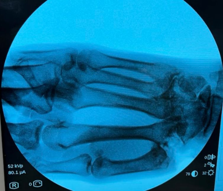
Figure 1: Intraoperative Fluoroscopy AP View of the Right Hand demonstrating complete excision of the trapezium bone

Figure 2: Intraoperative examination of complete removal of trapezium
Meanwhile, a gracilis cadaveric allograft is prepared on the back table. Utilizing 3-0 Vicryl suture, the graft is formed into an “anchovy” shape utilizing the ribbon technique where successive 1 cm lengths of the tendon are folded over the needle to form a structure which resembles a ribbon candy. This is then tightened down and secured with sutures to form a circular “anchovy” shape (Figure 3). This graft is inserted into the joint space as an interposition material to cushion and prevent bone-on-bone contact (Figure 4). Gelfoam is placed over the graft to assist with hemostasis. The periosteum and surrounding soft tissue are carefully closed with 4-0 Vicryl sutures to secure the graft in place. The extensor pollicis longus (EPL) tendon is examined to ensure it was not inadvertently tethered.
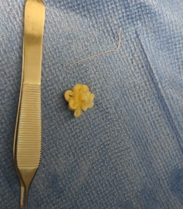
Figure 3: Gracilis Tendon prepared in an “Anchovy” shape
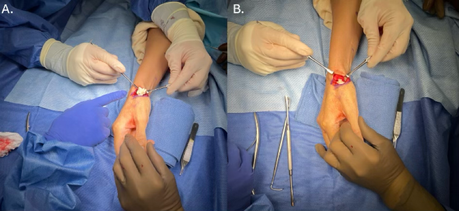
Figure 4: Intraoperative insertion of interpositional material; A: Placement of cadaveric “anchovy” shaped tendon into space; B: Snug fit of implanted graft with (not pictured) gelfoam overtop for hemostasis
A C-wire is then drilled from the proximal phalanx into the scaphoid to serve as an internal brace, stabilizing the repair. Proper placement of the wire is confirmed with X-ray (Figure 5). The wire is trimmed to an appropriate length, and a Jurgan ball is placed to protect the exposed end.

Figure 5: Intraoperative fluoroscopic imaging showing a PA view of the right hand with appropriate placement of C-wire through graft
The wound is copiously irrigated, and hemostasis is achieved. Skin edges are reapproximated with nylon sutures. Local anesthetic is injected into the carpal tunnel to target the median nerve, as well as over the superficial branch of the radial nerve for postoperative analgesia. Sterile dressings are applied, followed by a volar thumb spica splint. After releasing the tourniquet, brisk capillary refill is confirmed.
Postoperative Management
Postoperative outcomes and complications were recorded for up to 12 months following surgery, using data from a single hand surgeon’s database. Key outcomes included patient satisfaction, thumb disability scores, subjective assessments of pain and function, and the proportion of patients able to return to work.
Patients were monitored at regular intervals postoperatively. At the 2-week follow-up, the incisions had healed without complications. The internal brace wire was removed at 4 weeks, and patients began hand therapy focusing on range of motion exercises. By 2 months, most patients transitioned out of formal occupational therapy (OT), continuing with home exercises. At this point, the majority of patients demonstrated the ability to fully close their fists, grasp objects, and fully extend their fingers without difficulty, with most also returning to full work duties.
At 4 months postoperative, patients reported significant function compared to their preoperative state. Pain was markedly reduced, and patients noted improved capabilities in daily tasks. No difficulties were reported in using the operative hand for daily tasks. At this stage, patients reported no limitations in the Work Module of the DASH (Disabilities of the Arm, Shoulder, and Hand) score, achieving a perfect score of 0. Most returned to work without restrictions.
Follow-up typically continued until the 9-month mark, after which patients transitioned to as-needed visits with the clinic. In our small cohort, there were no significant adverse events reported.
Pearls and Pitfalls
Meticulous identification and protection of the SBRN is paramount when using a dorsal approach to the CMC joint. Gently mobilizing the SBRN branches and careful soft tissue retraction minimizes the risk of neuritis and permanent sensory complications. Be aware that the SBRN branches can traverse the surgical field at highly variable angles.13
Ensure complete excision of all trapezial bone, as leaving residual osteophytes particularly on the ulnar aspect or at the scaphoid-trapezium-trapezoid articulation creates persistent pain and impingement risk. Use intraoperative fluoroscopy or direct visualization to confirm complete removal of all diseased bones, including marginal osteophytes and any ulnar-sided remnants that could become pain generators postoperatively.
Carefully examine the extensor pollicis longus (EPL) tendon after graft placement to ensure it has not been inadvertently tethered or compressed. Entrapment by sutures, scar tissue, or graft impingement prevents thumb interphalangeal extension and significantly compromises opposition and pinch function, potentially necessitating revision surgery.
Complications
There were no complications reported by the patients in this series. Complications could include infection, complex regional pain syndrome, paresthesias, scar formation, delayed wound healing, metacarpal subsidence, and swelling.14
Discussion
This report describes the use of a gracilis tendon allograft for interposition following trapeziectomy. Patients in this series chose allograft to avoid donor-site morbidity and an additional incision. Autologous tendon harvest lengthens operative time, increases postoperative pain, and carries a risk of donor-site complications (hematoma, neuroma, adhesions, or weakness). Complete FCR harvest can reduce wrist flexion strength and endurance, which may affect recovery and overall satisfaction. Allografts avoid these concerns and are available in abundant supply. Tissue banks operate under federal regulation and perform extensive donor screening for infection and sterilization of processed tissue to minimize risk.15
Allografts avoid these concerns and are available in abundant supply. Tissue banks operate under federal regulation and perform extensive donor screening for infection and sterilizing processed tissues to minimize risk.16 Despite stringent protocols, contamination from bacteria such as Pseudomonas aeruginosa or Staphylococcus aureus remains possible, though rare.17,18 Viral and prion transmission is theoretically possible but is highly uncommon with nucleic acid testing. Immune rejection is another theoretical, though infrequent, risk.19
Literature confirms favorable outcomes using allografts in basal joint arthroplasty. Marks et al.20 found human dermal collagen templates yielded equivalent one-year outcomes to FCR autografts when evaluated by MHQ and DASH scores. McCullough et al.21 reported similar improvements in grip and pinch strength using meniscal allografts with less metacarpal subsidence. Kokkalis et al.16 observed that 89 patients reconstructed with acellular dermal allografts had reduced pain and improved grip and key pinch compared to baseline. These findings underscore that allograft materials can replicate autograft performance while simplifying surgery.
Cadaveric tissue offers distinct advantages: it avoids the harvest-related morbidity of autograft and the foreign-body reactions associated with synthetic implants. The gracilis tendon, in particular, provides excellent pliability and volume that make it ideal for interpositional cushioning in basal joint arthroplasty. Its rounded structure delivers durable bulk while maintaining tendinous consistency for stability, without the inflammatory responses often seen with synthetic materials.
Gracilis allograft suspension-interposition arthroplasty yields several key benefits, including reduced operative and tourniquet times, elimination of donor-site pain and scarring, fewer incisions with faster rehabilitation, and avoidance of the foreign-body reactions associated with synthetic implants. We did not perform a cost analysis, so any economic advantage over implants or autograft remains unverified. This case series is limited by its small sample size, lack of a control group, and short 12-month follow-up. Future studies with larger cohorts, longer-term radiographic evaluation of metacarpal subsidence and stability, and validated patient-reported outcomes are essential to confirm whether gracilis allografts outperform established methods like isolated trapeziectomy or flexor carpi radialis interposition.
Conclusion
Gracilis tendon allograft suspensioninterposition arthroplasty appears to be a safe, efficient, and effective option for managing advanced thumb CMC arthritis. It eliminates donor morbidity, simplifies the procedure, reduces recovery time, and avoids implant-related complications. While preliminary, these results support further investigation into gracilis allografts as a viable alternative graft source in basal joint arthroplasty.
Acknowledgements: None
Ethics Approval and Consent to Participate: This case report does not contain any animal subjects. This case report did not require institutional ethical approval. Written informed consent was obtained from patients for publication of the case report and respective images.
Funding: The authors declare no funding was received for this technique guide
References
- Hartnett DA, Milner JD, DeFroda SF. Osteoarthritis in the upper extremity. Am J Med. 2023;136(5):415-421. doi:10.1016/j.amjmed.2023.01.025
- Weiss AC, Goodman AD. Thumb basal joint arthritis. J Am Acad Orthop Surg. 2018;26(16):562-571. doi:10.5435/JAAOS-D-17-00374
- Athlani L, De Almeida YK, Martins A, Seaourt AC, Dap F. Thumb basal joint arthritis in 2023. Orthop Traumatol Surg Res. 2024;110(1S):103772. doi:10.1016/j.otsr.2023.103772
- Yoon AP, Hutton DW, Chung KC. Cost-effectiveness of surgical treatment of thumb carpometacarpal joint arthritis: a value of information study. Cost Eff Resour Alloc. 2023;21(1):28. doi:10.1186/s12962-023-00438-8
- Polatsch DB, Paksima N. Basal joint arthritis: diagnosis and treatment. Bull NYU Hosp Jt Dis. 2006;64(3-4):178-184.
- Spaans AJ, Weijns ME, Braakenburg A, van Minnen LP, Mink van der Molen AB. Partial trapeziectomy and interposition of fascia lata allograft in the operative treatment of thumb base osteoarthritis. J Plast Surg Hand Surg. 2016;50(1):35-39. doi:10.3109/2000656X.2015.1069744
- Nordin JS, Olsson O, Lunsjö K. The gracilis tendon autograft is a safe choice for orthopedic reconstructive procedures: a consecutive case series studying the effects of tendon harvesting. BMC Musculoskelet Disord. 2019;20(1):138. doi:10.1186/s12891-019-2520-5
- Wolock BS, Moore JR, Weiland AJ. Arthritis of the basal joint of the thumb. A critical analysis of treatment options. J Arthroplasty. 1989;4(1):65-78. doi:10.1016/s0883-5403(89)80055-5
- Nichols DS, Oberhofer HM, Chim H. Anatomy and biomechanics of the thumb carpometacarpal joint. Hand Clin. 2022;38(2):129-139. doi:10.1016/j.hcl.2021.11.001
- Carlson Strother C, Pulos N. Vascular supply of the wrist. Hand Clin. 2022;38(4):377-384. doi:10.1016/j.hcl.2022.03.001
- Jongen IC, Nieuwdorp NJ, Hundepool CA, Van Gelder FS, Schutter AM, Zuidam JM. Ligament reconstruction in thumb carpometacarpal joint instability: a systematic review. JPRAS Open. 2024;39:237-248. doi:10.1016/j.jpra.2024.01.001
- Rezzadeh K, Rossi K, Trerotola CC, Shah A. First carpometacarpal joint denervation: a systematic review. J Hand Surg Am. 2022;47(8):793.e1-793.e8. doi:10.1016/j.jhsa.2021.07.020
- Hemmi S, Kurokawa K, Nagai T, et al. Anatomical variations of the superficial branch of the radial nerve and the dorsal branch of the ulnar nerve: a detailed electrophysiological study. Muscle Nerve. 2021;63(6):913-918. doi:10.1002/mus.27221
- Naram A, Lyons K, Rothkopf DM, et al. Increased complications in trapeziectomy with ligament reconstruction and tendon interposition compared with trapeziectomy alone. Hand (N Y). Mar 2016;11(1):78-82. doi:10.1177/1558944715617215
- Kokkalis ZT, Zanaros G, Weiser RW, Sotereanos DG. Trapezium resection with suspension and interposition arthroplasty using acellular dermal allograft for thumb carpometacarpal arthritis. J Hand Surg Am. 2009;34(6):1029-1036.
- Getgood A, Bollen S. What tissue bankers should know about the use of allograft tendons and cartilage in orthopaedics. Cell Tissue Bank. 2010;11(1):87-97. doi:10.1007/s10561-009-9129-0 doi:10.1016/j.jhsa.2009.03.001
- Beer AJ, Tauro TM, Redondo ML, Christian DR, Cole BJ, Frank RM. Use of allografts in orthopaedic surgery: safety, procurement, storage, and outcomes. Orthop J Sports Med. 2019;7(12):2325967119891435. doi:10.1177/2325967119891435
- Harner CD, Lo MY. Future of Allografts in Sports Medicine. Clin Sports Med. 2009;28(2):327-340. doi:10.1016/j.csm.2008.10.010
- Betz RR. Limitations of autograft and allograft: new synthetic solutions. Orthopedics. 2002;25(5 Suppl):S561-S570. doi:10.3928/0147-7447-20020502-04
- Marks M, Hensler S, Wehrli M, Scheibler AG, Schindele S, Herren DB. Trapeziectomy with suspension-interposition arthroplasty for thumb carpometacarpal osteoarthritis: a randomized controlled trial comparing the use of allograft versus flexor carpi radialis tendon. J Hand Surg Am. 2017;42(12):978-986. doi:10.1016/j.jhsa.2017.07.023
- McCullough MC, Minasian R, Tanabe K, Rodriguez S, Kulber D. Functional outcomes for basilar joint arthroplasty with meniscus allograft compared with trapeziectomy alone. Hand (N Y). 2023;18(1):89-97. doi:10.1177/1558944721999730