
Hunter Hitchens B.S.1; Saliha Ahmad M.S.1; William H. Fang D.O.2; Mitchell Tingey M.D.2; Kevin Mo D.O.2; Carl Williams M.D.2
1Touro University Nevada1, Department of Orthopaedic Surgery
2Valley Hospital Medical Center, Las Vegas, NV
Abstract
Background
Contrast media (CM) extravasation is a rare complication of imaging procedures, usually benign but occasionally progressing to acute compartment syndrome (ACS), a limb-threatening emergency requiring prompt surgical decompression.
Case Presentation
A 47-year-old woman with pancreatic cancer and type I diabetes developed severe left hand swelling, pain, and numbness following a CT angiogram. Imaging revealed extensive CM extravasation throughout the forearm and wrist. Emergent fasciotomy revealed tense, edematous compartments with expression of extravasated contrast, consistent with ACS; compartment pressures were not formally measured. Staged wound management included negative pressure wound therapy, Restrata synthetic sheath application, and autologous skin grafting. A return to the OR at 48 hours was delayed due to the patient suffering an acute anxiety attack, however the patient ultimately regained significant motor and sensory function.
Discussion and Conclusion
This case underscores the potential for ACS following CM extravasation and the critical importance of early recognition. Literature suggests no universal consensus on optimal fasciotomy closure, highlighting the need for comparative studies to establish evidence-based guidelines.
Keywords: Contrast Media Extravasation; Acute Compartment Syndrome; Fasciotomy Closure Techniques; Negative Pressure Wound Therapy; Skin Grafting; Case Report
Introduction
Contrast media (CM) extravasation refers to the leakage of intravascularly injected contrast into the surrounding soft tissues during imaging procedures [1, 2, 3]. This complication is rare, occurring in approximately 0.1% to 1.2% of contrast injections [1, 2, 3, 4, 5]. In most cases, CM extravasation is generally associated with mild self-limited symptoms such as transient pain, swelling, and erythema. These typically resolve with conservative management, including limb elevation, cold compresses, and observation [1, 2, 4, 5]. However, in rare instances, extravasation of large volumes of hyperosmolar contrast under high pressure can result in acute compartment syndrome (ACS) [1, 2, 3, 6, 7, 8].
Several factors contribute to the likelihood of extravasation. Higher flow rates used in CT angiography increases intraluminal pressure and shear stress on venous walls, thereby elevating the likelihood of extravasation [2, 9, 10, 11]. Patient-specific factors have also been implicated, including sex, inpatient status, and the location of the IV insertion [5, 9, 11]. Females are at higher risk than males to experience CM extravasation, possibly due to smaller vessel caliber [9, 11]. Inpatients often have IVs placed by non-radiology staff, sometimes hours before contrast administration, increasing the chance of a catheter dislodgement or migration [4, 9, 11]. Furthermore, there are higher rates of CM extravasation seen in IVs placed on the dorsum of the hand, where the veins are smaller and more fragile than those in the antecubital fossa [1, 3, 4, 5, 7, 9].
ACS is a surgical emergency in which elevated compartment pressure compromises perfusion and threatens tissue viability [2, 3, 8, 12]. Without timely decompression, rising compartment pressure can progress to muscle necrosis, permanent nerve damage, and limb loss [3, 5, 8, 13]. Clinical suspicion for ACS should be raised in the presence of the classic “5 P’s”: pain, pallor, paresthesia, paralysis, and pulselessness [8, 12, 13]. Early recognition of these signs is critical, as ACS requires urgent surgical fasciotomy to relieve pressure and restore circulation [3, 5, 8, 12, 13].
Crucial steps of a successful fasciotomy include an extensile incision, complete fascial release of all affected compartments, preservation of critical structures, debridement of necrotic tissue, and delayed wound closure [1, 6, 8, 14]. Postoperative wound management is equally important and must account for factors such as, tissue viability, wound size, timing of closure, and patient comorbidities. Negative pressure wound therapy (NPWT), or vacuum-assisted closure following a fasciotomy for ACS, is designed to prepare the wound for healing and secondary closure [8]. The technique applies sub-atmospheric pressure to the wound via sealed dressing, thereby facilitating continuous exudate removal, reducing edema, and improving local perfusion [8]. In fasciotomy wounds, particularly those caused by CM extravasation, NPWT stabilizes the wound environment until definitive closure is feasible [13]. It maintains a moist, sterile environment while preventing further tissue distension and increases in compartment pressure [11]. NPWT enhances graft integration and reduces infection rates when compared to conventional dressings [12].
In this case report, we present a rare instance of ACS of the upper extremity following CM extravasation. The case is notable not only for the unusual etiology of ACS, but also for the complex, staged surgical management that followed. Over five weeks, the patient underwent multiple operative interventions to restore perfusion, promote wound healing, and preserve limb function. This case highlights the use of various wound closure techniques, including NPWT, synthetic wound matrices, and autologous skin grafting to manage the fasciotomy site. We aim to illustrate the challenges of postoperative wound management in ACS and to contribute to the growing discussion on optimizing closure strategies in complex soft tissue injuries.
Case Presentation
A 47-year-old female with a past medical history of pancreatic cancer and type 1 diabetes mellitus presented to the emergency department (ED) in 2025, at approximately 1:00 AM with altered mental status, abdominal pain, nausea, and vomiting.
On arrival, the patient was somnolent, intermittently agitated, and oriented only to self and situation. Collateral history obtained from her husband revealed she was last known to be at her neurological baseline approximately 11 hours prior to presentation. Emergency medical services reported a blood glucose level in the 400s en route and noted peaked T waves on the prehospital electrocardiogram. (Table 1)
Table 1. Initial Laboratory Evaluation
| Lab Test | Lab Value | Interpretation |
| Blood Glucose | 400 → 420 mg/dL | Elevated; no evidence of diabetic ketoacidosis |
| Lactic Acid | 4 → 6 mmol/L | Rising despite fluid resuscitation |
| Ethanol | Negative | N/A |
| Troponin | Within normal limits | No myocardial injury |
| WBC Count | Within normal limits | No leukocytosis |
| Liver Function Tests | Within normal limits | No hepatic dysfunction |
In the ED, the patient’s blood pressure responded well to intravenous labetalol. Pain and agitation were managed with IV morphine and IV lorazepam (Ativan), respectively. A non-contrast CT scan of the head revealed no acute intracranial pathology. Due to multiple symptoms and severe hypertension with unclear history, a CT angiogram of the chest, abdomen, and pelvis was performed to evaluate for aortic dissection. While no aortic aneurysm was identified, the radiologist could not definitively exclude dissection. Following initial stabilization, the patient’s mental status improved, but due to the rising lactic acid levels, she was admitted for further management.
Overnight, the patient developed increasing pain, swelling, and paresthesia in the left upper extremity (LUE). Hand surgery was urgently consulted due to concern for ACS. Physical examination revealed blistering at the IV site on the wrist, diffuse swelling, marked tenderness, and absence of active wrist movement. The patient reported increasing numbness and tingling in the fingertips, with diminished sensation and radial and ulnar pulses weaker than on the contralateral side. A non-contrast CT scan of the LUE confirmed a large-volume contrast extravasation, extending from the dorsal radial elbow through the forearm and to the dorsum of the hand. (Figure 1 and 2)
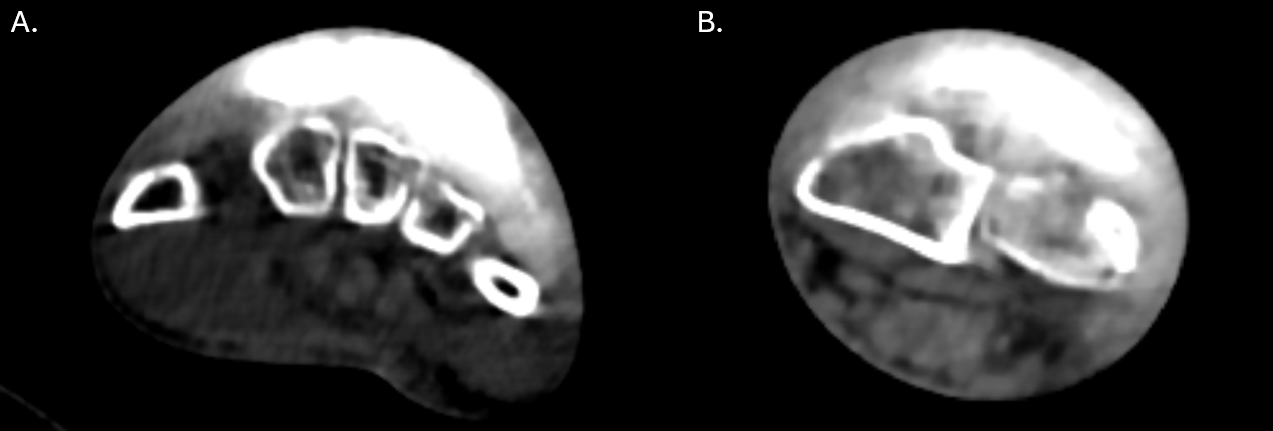
Figure 1. (a) CT Axial Left Hand showing a large-volume radiopaque contrast extravasation dorsally; (b) CT Axial Left Proximal Aspect of the Distal Radio-Ulnar joint showing a large-volume radiopaque contrast extravasation dorsally.

Figure 2. (a) CT Coronal Left arm showing a large-volume radiopaque contrast extravasation ulnarly; (b) CT Sagittal Left Elbow showing a large-volume radiopaque contrast extravasation dorsally.
Given the physical exam and radiologic findings were consistent with ACS, the decision was made to take the patient to the operating room for an emergent fasciotomy of the LUE. Prior to surgical preparation, no palpable or Doppler-identifiable pulses were present in the affected extremity. All compartments of the forearm and hand were opened and released, including the carpal tunnel as part of the volar approach. (Figure 3) No frank necrosis was noted, though the tissues were tense and edematous. A large amount of clear fluid was expressed in the incisions, consistent with IV contrast. The wounds were irrigated with copious amounts of normal saline. Given the degree of tissue edema, no primary closure was attempted. The wounds were loosely packed with sterile dressings, and 2 NPWT devices were applied to all fasciotomy sites.
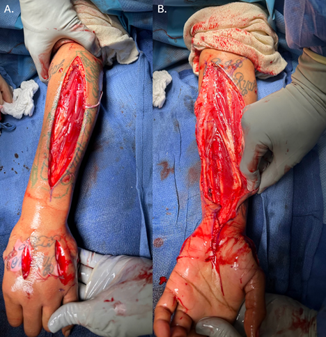
Figure 3. (a) Dorsal fasciotomy incisions showing complete decompression of the forearm compartments.; (b) Volar forearm fasciotomy showing release of the flexor compartment with viable muscle.
In the immediate postoperative period following fasciotomy of the LUE, intraoperative cultures demonstrated bacterial growth of Staphylococcus aureus, including methicillin-resistant S. aureus (MRSA). The infectious diseases team was consulted, and empiric antibiotic therapy was initiated with intravenous vancomycin, daptomycin, and ampicillin-sulbactam (Unasyn).
On postoperative day 1, the patient was noted to be doing clinically well. The NPWT device was in place and functioning appropriately. (Figure 4). Pain was well-controlled with standard analgesic protocols. Neurological exam revealed motor strength of 2/5 with some muscle firing; however, sensation remained absent. Pulses were strong and symmetrical at 2+ bilaterally.
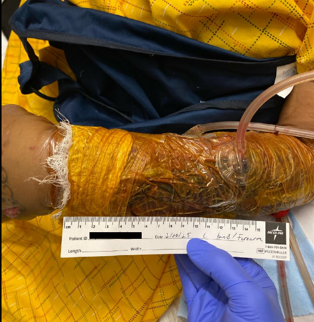
Figure 4. Wound VAC placement following fasciotomy surgery
The surgical team initially planned a return to the operating room in two days for further irrigation and debridement, and primary wound closure. However, this procedure was delayed due to an incident in the preoperative area where the patient became acutely agitated and anxious upon encountering a staff member, likely due to underlying psychiatric comorbidities. In light of this behavioral episode, the procedure was subsequently rescheduled.
Second Operation
At the time of the second surgery, a 12 × 2 cm dorsal wound area was closed primarily using a combination of interrupted 3-0 nylon sutures and mattress suturing technique. The residual open area, measuring 10 × 6 cm, was covered with a meshed Restrata Wound Matrix (RVM) sheath and secured using 3-0 Vicryl sutures. (Figure 5) A similar closure technique was performed dorsally with another RVM application (10 × 3 cm), which was meshed and tacked centrally with 3-0 Vicryl to prevent tenting. An NPWT device was reapplied postoperatively. Plans were made for future skin grafting of the remaining exposed areas.
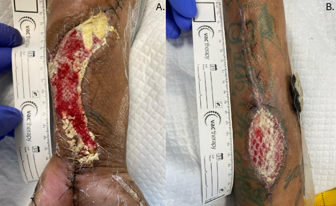
Figure 5. (a) Volar wound pictures POD 5 from initial debridement after Wound VAC removal. Skin was loosely approximated with 2-0 nylon sutures.; (b) Dorsal wound pictures POD 5 from initial debridement after Wound VAC removal. Skin was loosely approximated with 2-0 nylon sutures.
Subsequent Recovery and Interventions
Approximately 10 days after the initial fasciotomy, the patient began to report gradual return of sensation in the affected hand. She remained on intravenous antibiotics, and continued with NPWT and comprehensive medical management. Occupational and physical therapy teams were actively engaged during her hospitalization, focusing on restoring hand mobility and function.
Despite scheduled plans for further surgical interventions, multiple procedures were postponed due to recurrent psychiatric disturbances and episodes of agitation. Ultimately, the final surgical intervention was performed on March 28, 2025, consisting of debridement and skin grafting.
Third Operation
A full-thickness skin graft (FTSG) was harvested from the right hip for application to the volar aspect of the left hand. The initial defect measured 10 × 6 cm, which was debrided to 10.5 × 6.5 × 0.7 cm. The graft was meshed in a 1:1.5 ratio, cut to fit the defect, and secured with surgical staples. Concurrently, the dorsum of the left forearm, with an initial defect measuring 16 × 6 cm, was debrided to an area of 6.5 × 6.5 × 0.7 cm and received a split-thickness skin graft (STSG), which was also trimmed, contoured, and stapled in place. Adaptic, topical thrombin, and OpSite dressings were applied over both sites, followed by placement of a wound VAC over the donor site on the right leg. The patient tolerated the procedure well and demonstrated good capillary refill postoperatively.
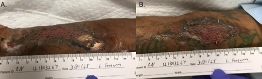
Figure 6. (a) Volar wound pictures POD 2 after full-thickness skin graft application demonstrating progressive wound healing with improved granulation tissue.; (b) Dorsal wound pictures POD 2 after split-thickness skin graft application demonstrating progressive wound healing with improved granulation tissue.
Follow-Up and Functional Recovery
The patient was discharged three days following the final surgical procedure. She was evaluated in the clinic two weeks later; the grafts were noted to be well integrated, and surgical staples were removed. At her one-month follow-up, she demonstrated further improvement in sensation and range of motion. By the three-month mark, she reported approximately 60% resolution of her initial symptoms, with continued progressive healing of the hand.
At six months postoperatively, the patient reported approximately 80% resolution of her symptoms and had regained significant functional use of the hand. She was planning to relocate to California and intended to continue care with a new hand surgeon in her new location.
Discussion
This case highlights ACS of the LUE, a rare and serious complication of CM extravasation. Several cases of ACS developing after high-pressure contrast injections during imaging studies have been documented [1, 3, 6, 7, 13]. Two consistent factors across these reports are extravasations involving large volumes (over 50-100 mL) and the use of power-injected CM through a small dorsal hand vein [1, 2, 3, 5, 7]. Additionally, predisposing factors and comorbidities increase the patient’s risk of injury exacerbation. Relevant examples in this case are chemotherapy-induced vascular fragility and diabetes-related microvascular impairment [3].
Once ACS was recognized, the patient underwent emergent fasciotomies, consistent with past reported cases in which prompt surgical decompression is recommended to prevent irreversible tissue damage [1, 6, 7, 16]. A series of interventions was required to completely close the wounds from the fasciotomies, as is typical following this procedure. During the first postoperative period, the surgeons applied NPWT to reduce edema, promote granulation tissue formation, and allow infected tissues time to respond to antibiotic therapy. The wound VAC system maintained continuous suction, allowing removal of excess exudate whilst reducing tissue tension at fasciotomy sites. In subsequent surgeries, the LUE was first treated with a series of primary closures using interrupted and mattress sutures, combined with synthetic wound sheaths (Restrata) to bridge remaining soft tissue gaps. These scaffolds support re-epithelialization, minimize inappropriate wound contracture, and maintain an environment conducive to optimal tissue repair. The final wound closure was done using both a FTSG and STSG that were harvested from the right hip and thigh donor sites. The FTSG provided durable coverage and improved aesthetic outcomes for high-stress areas demanding greater dermal integrity, including the volar wrist and hand. In contrast, the STSG was used over larger forearm regions to improve graft take through increased vascularization.
Throughout the case, the patient experienced several acute psychiatric episodes between surgical interventions. These events contributed to repeated postponements of debridement and skin grafting, thereby prolonging the overall healing timeline and postoperative recovery. Nonetheless, this sequence of surgical procedures was well tolerated and resulted in adequate functional restoration.
Conclusion
CM extravasation–induced compartment syndrome is a rare but serious complication requiring rapid diagnosis and urgent fasciotomy to prevent permanent tissue damage. Optimal recovery depends not only on timely decompression but also on meticulous postoperative wound management. The optimal closure method remains uncertain and likely varies with patient and wound characteristics. Further comparative studies are needed to establish standardized, evidence-based guidelines for fasciotomy closure that balance efficacy, safety, and resource considerations.
Informed Consent
Patient consent was obtained to use the information in this case report for educational purposes.
Funding
This research did not receive any specific grant from funding agencies in the public, commercial, or not-for-profit sectors.
Abbreviations
| CM | Contrast media |
| ACS | Acute compartment syndrome |
| NPWT | Negative pressure wound therapy |
| ED | Emergency Department |
| LUE | Left upper extremity |
| MRSA | Methicillin-resistant S. aureus |
| FTSG | Full-thickness skin graft |
| STSG | Split-thickness skin graft |
| RWM | Restrata Wound Matrix |
References
- Diop B, Soh GT, Nde AFT, Sarr N, Ndoye AY, Sane JC. Hand compartment syndrome secondary to contrast media extrava-sation: A case report. Int J Surg Case Rep. 2022;98:107595. doi:10.1016/j.ijscr.2022.107595
- Raveendran S, Rajendra Benny K, Monica S, Pallapati SR, Keshava SN, Thomas BP. Multiple Stab Incisions and Evacuation Technique for Contrast Extravasation of the Hand and Forearm. J Hand Surg Am. 2019;44(1):71.e1-71.e5. doi:10.1016/j.jhsa.2018.08.009
- van Veelen NM, Link BC, Donner G, Babst R, Beeres FJP. Compartment syndrome of the forearm caused by contrast medium extravasation: A case report and review of the literature. Clin Imaging. 2020;61:58-61. doi:10.1016/j.clinimag.2020.01.013
- Dykes TM, Bhargavan-Chatfield M, Dyer RB. Intravenous contrast extravasation during CT: a national data registry and practice quality improvement initiative. J Am Coll Radiol. 2015;12(2):183-191. doi:10.1016/j.jacr.2014.07.021
- Vinod KV, Shravan R, Shrivarthan R, Radhakrishna P, Dutta TK. Acute compartment syndrome of hand resulting from ra-diographic contrast iohexol extravasation. J Pharmacol Pharmacother. 2016;7(1):44-47. doi:10.4103/0976-500X.179353
- Savage SA, Fox C, Goldie SJ. Compartment syndrome of the hand following CT contrast extravasation. ANZ Journal of Sur-gery. Published online November 18, 2022. doi:10.1111/ans.18155
- Stavrakakis IM, Daskalakis II, Detsis EPS, Karagianni CA, Papantonaki SN, Katsafarou MS. Hand compartment syndrome as a result of intravenous contrast extravasation. Oxf Med Case Reports. 2018;2018(12):omy098. Published 2018 Nov 21. doi:10.1093/omcr/omy098
- Vyskocil J, Sheu J, Telehowski P. Hand and Forearm Compartment Syndrome Secondary to Intravenous Infiltration. J Orthop Case Rep. 2024;14(11):92-96. doi:10.13107/jocr.2024.v14.i11.4926
- Heshmatzadeh Behzadi A, Farooq Z, Newhouse JH, Prince MR. MRI and CT contrast media extravasation: A systematic review. Medicine (Baltimore). 2018;97(9):e0055. doi:10.1097/MD.0000000000010055
- Sakellariou S, Li W, Paul MC, Roditi G. Rôle of contrast media viscosity in altering vessel wall shear stress and relation to the risk of contrast extravasations. Med Eng Phys. 2016;38(12):1426-1433. doi:10.1016/j.medengphy.2016.09.016
- Hwang EJ, Shin CI, Choi YH, Park CM. Frequency, outcome, and risk factors of contrast media extravasation in 142,651 in-travenous contrast-enhanced CT scans. Eur Radiol. 2018;28(12):5368-5375. doi:10.1007/s00330-018-5507-y
- Cianci M, Nicolosi B, Pinzauti E, Pancani S. Paediatric acute compartment syndrome of the limbs due to fluid extravasation: a case study. Wounds International. 2023;14(4).
- Tirrell AR, Nigam M, Hung RW. Isolated Compartment Syndrome of the Hand After Intravenous Doxycycline Infiltration Injury. J Hand Surg Glob Online. 2022;4(4):239-243. Published 2022 Jan 10. doi:10.1016/j.jhsg.2021.12.002
- Donaldson J, Haddad B, Khan WS. The pathophysiology, diagnosis and current management of acute compartment syndrome. Open Orthop J. 2014;8:185-193. Published 2014 Jun 27. doi:10.2174/1874325001408010185
- Igoumenou VG, Kokkalis ZT, Mavrogenis AF. Fasciotomy Wound Management. Compartment Syndrome. Published online 2019:83-95. doi:https://doi.org/10.1007/978-3-030-22331-1_9
- Barber CM, Fahrenkopf MP, Adams NS, Kelpin JP, Krebiehl JR. Compartment Syndrome of the Hand Secondary to Intravenous Extravasation. Eplasty. 2018;18:ic17. Published 2018 Sep 24
- Jauregui JJ, Yarmis SJ, Tsai J, Onuoha KO, Illical E, Paulino CB. Fasciotomy closure techniques. J Orthop Surg (Hong Kong). 2017;25(1):2309499016684724. doi:10.1177/2309499016684724
- Medina C, Spears J, Mitra A. The use of an innovative device for wound closure after upper extremity fasciotomy. Hand (N Y). 2008;3(2):146-151. doi:10.1007/s11552-007-9082-y
- MacEwan MR, MacEwan S, Kovacs TR, Batts J. What Makes the Optimal Wound Healing Material? A Review of Current Science and Introduction of a Synthetic Nanofabricated Wound Care Scaffold. Cureus. 2017;9(10):e1736. Published 2017 Oct 2. doi:10.7759/cureus.1736
- MacEwan M, Jeng L, Kovács T, Sallade E. Clinical Application of Bioresorbable, Synthetic, Electrospun Matrix in Wound Healing. Bioengineering (Basel). 2022;10(1):9. Published 2022 Dec 21. doi:10.3390/bioengineering10010009
- Kohlhauser M, Luze H, Nischwitz SP, Kamolz LP. Historical Evolution of Skin Grafting-A Journey through Time. Medicina (Kaunas). 2021;57(4):348. Published 2021 Apr 5. doi:10.3390/medicina57040348
- Elseth A, Nunez Lopez O: Wound Grafts, in StatPearls. Treasure Island, FL, StatPearls, 2025.
- Putri IL, Sindhu FC, Aisyah IF, Pramanasari R, Wungu CDK. Comparison of combination skin substitutes and skin grafts versus skin grafts only for treating wounds measured by Vancouver Scar Scale: A comprehensive meta-analysis. SAGE Open Med. 2024;12:20503121241266342. Published 2024 Aug 12. doi:10.1177/20503121241266342
- Govaert GA, van Helden S. Ty-raps in trauma: a novel closing technique of extremity fasciotomy wounds. J Trauma. 2010;69(4):972-975. doi:10.1097/TA.0b013e3181f2d9d3
- Capo J, Liporace F, Yingling JM, et al. Pressure reducing skin pie-crusting in extremity trauma: An in-vitro biomechanical study and human case series. Injury. 2020;51(6):1266-1270. doi:10.1016/j.injury.2020.03.008
- Kazaleh M, Prange EJ, Madbak FG. Dermal fenestration ‘pie crust’ technique for difficult fasciotomy closures. Trauma Surg Acute Care Open. 2023;8(1):e001212. Published 2023 Aug 28. doi:10.1136/tsaco-2023-001212
- Brown LR, Rentea RM. Temporary Abdominal Closure Techniques. In: StatPearls. Treasure Island (FL): StatPearls Publishing; February 24, 2024.
- Çelik M, Öztürk V, Çatal T, Bayrak A, Duramaz A, Bilgili MG. The efficacy of the Bogota Bag technique for wound closure in limb fasciotomy patients: a prospective cohort study. Int Orthop. 2024;48(11):2971-2978. doi:10.1007/s00264-024-06292-4