Banks J, Nnisar A, Harper T, Banks J
Lincoln Memorial University – DeBusk College of Osteopathic Medicine, Knoxville, Tennessee, United states of America
Intro: The tibia is the main weight-bearing bone of the leg, with the fibula supporting this function and providing an attachment point for various muscles. We conducted a three-dimensional analysis to quantify cross-sectional geometry characteristics of paired tibiae and fibulae to determine if mechanical stiffness is correlated between these bones. We hypothesize that the fibula compensates for resistance to bending in areas where the tibia is weak and/or thin. This information can help to explain certain breakage patterns that are commonly seen by orthopedic physicians.
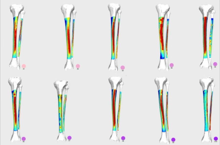
Methods: The CT data from 10 deceased individuals were obtained with permission from the New Mexico Descendent Image Database. CT data from the left leg of each donor were cropped, and the cortical bone was segmented to generate digitized periosteal and endosteal surfaces of both the tibia and fibula. We used these to quantify cross-sectional parameters across 30 equidistant slices taken from the central diaphysis. This data was then analyzed using the Morphomap cross-sectional geometry package in R. Non-parametric correlations were measured between resistances to mediolateral and anteroposterior bending, longitudinal twisting, and mean diaphyseal thickness and cortical area.
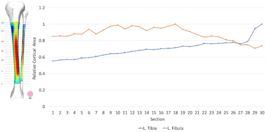
Results: We found that cortical area was positively correlated in both bones with body mass and height, and negatively correlated with age. Comparing the cross-sectional properties of coplanar sections of the tibia and fibula, we found that all second moment of area measurements in the tibia were negatively correlated with cortical area and mean thickness of the fibula. Resistances to mediolateral bending and axial torsion were also found to be inversely proportional. Generally, the diaphyseal cortical bone of the tibia becomes thinner distally, while the diaphysis of the fibula is thickest in the middle and is at its thinnest at its superior and inferior ends.
Conclusion: These results support the hypothesis that the fibula partially compensates for the reduced bending resistance of the tibia in regions where the tibial cortical bone is particularly thin and/or weak. These data inform functional hypotheses of the fibula and can help explain the mechanism of certain breakage patterns in fractures involving the distal lower limb.