
Christian Allen, DO1; Andres Toledo, DO1; Ryan Dennett, OMS 20282; John Harker, DO1; Richard Crank, DO1
1HCA Largo Florida/USF Morsani College of Medicine
2University of North Texas Texas College of Osteopathic Medicine
DOI: http://doi.org/10.70709/ezvkkk4dcmulbu
ABSTRACT
Background
This level IV evidence case report describes the surgical and wound management of chronic necrotizing fasciitis of the right lower extremity in a 73-year-old female after a gardening tool caused deep penetration into her posterior calf, which required circumferential removal of skin and subcutaneous fat along with multiple a fasciotomy, multiple irrigations and debridement’s, wound vacuums applications, and two rounds of application of biodegradable temporizing matrix. This case outlines a multistage process for treatment and management following radical removal of soft-tissue coverage for deep wound/compartment infection.
Methods
Patient presented with a 1cm opening to the lateral right midportion of her lower extremity with erythema and serous drainage. White blood cell count, erythrocyte sedimentation rate, and C-reactive protein were elevated. Computer tomography of the right lower extremity demonstrated multiple large abscess collections in subcutaneous tissues and in the posterior compartment of the lower extremity extending proximally to the mid-calf. The patient’s clinical findings, laboratory, and imaging evaluation led the patient to require a fasciotomy, multiple irrigations and debridement’s, wound vacuums applications, and two rounds of application of biodegradable temporizing matrix. Broad spectrum intravenous antibiotics were administered and tailored to intra-operative cultures. Clinical and functional outcomes were assessed at follow-up.
Results
Three months after the second application of biodegradable temporizing matrices, a physical exam demonstrated circumferential granulation tissue present about the right lower extremity with no evidence of active bleeding, drainage, or purulent material. The biodegradable temporizing matrix was adhered with granulation tissue formation. The proximal medial incision was closed, dry, and intact with scarring. The patient was neurovascularly intact with palpable pedal pulses and ambulating at will.
Conclusion
Management of necrotizing fasciitis in the setting of deep infections with multicompartment involvement remains complex, particularly in patients with delayed presentation, which allows the disease process to propagate. Accurate preoperative quantification of the infected area, thoughtful wound management, and careful consideration of surgical debridement are critical to optimize outcomes. This case demonstrates that a multistage strategy, combined with a biodegradable temporizing matrix, can restore soft-tissue coverage and function. This approach provides treatment of the infection without recurrent wound nonhealing. To our knowledge, this is the first report of a multistage surgical procedure for necrotizing fasciitis with extensive circumferential soft-tissue loss and the use of a biodegradable temporizing matrix, offering a potential treatment framework for similarly challenging presentations.
Keywords: Necrotizing Fasciitis, Application of Skin Graft, Biodegradable Temporizing Matrix, Complex Lower Extremity Wound, Multi-Compartment Involvement
Introduction
Necrotizing fasciitis (NF) is a rare, rapidly-progressive bacterial infection known to affect various fascial planes that, if left untreated, can progress from fascia and subcutaneous tissue to a secondary infection involving underlying soft tissues and muscles.6,16 These deep, penetrating infections result in extensive tissue necrosis; therefore, urgent surgical intervention is required to achieve adequate control of the infection source, limit tissue loss, and minimize the risk of amputation of the affected extremity.18
Early clinical diagnosis is a critical outcome determinant. However, considering that physical exams, imaging, and laboratory values are rarely pathognomonic, surgery remains the preferred method of treatment as it can not only improve outcomes but can provide a definitive diagnosis.2,12 Thus, the current management of NF involves the usage of broad-spectrum empirical antibiotics in conjunction with early surgical fasciotomy and aggressive debridement in a staged manner.12 While these therapeutic interventions have been shown to improve mortality rates, the extensive debridement that is involved results in associated impaired or delayed wound healing.7 Disruption of the local vasculature and the corresponding chronic inflammatory response creates an environment that increases the risk of healing complications.
Biodegradable Temporizing Matrix (BTM) is a potential solution to the problem orthopedist face for complex wound healing. BTM is a bi-layer synthetic skin composite consisting of a biodegradable polyurethane matrix foam covered with a nonbiodegradable polyurethane sealing membrane intended to mimic the dermis and epidermis, respectively.8,15 Its porous matrix supports cell migration and proliferation, promoting neoangiogenesis and vascularization while also functioning as a biodegradable polyurethane scaffold for the formation of a neodermis.14 The application process of this matrix involves a two-step procedure. The first stage is the integration phase, where the vascularized neodermis develops on a clean wound bed, followed by the sealing membrane being removed and a split-thickness skin graft being applied.9 A key difference between other biologic dermal substitutes and BTM is that it is fully synthetic, which avoids the risk of an associated immunological response or disease transmission while maintaining its structural integrity within an inflamed or contaminated wound environment.5
The purpose of this case report is to illustrate the presentation of a female with deep seeded necrotizing fasciitis with delayed presentation who developed significant loss of tissues during multiple irrigations and debridements, and to outline the staged surgical approach that was employed to restore soft tissue coverage.
Case Report
A 73-year-old female presented to our institution’s emergency department with a greater than 1 month wound to her right lower extremity. Patient stated she accidentally stabbed herself with a gardening tool and did not seek medical attention. She has been self-medicating with hydrogen peroxide until she noticed redness, swelling, skin sloughing, drainage from the lesion site, and mild pain during ambulation. The patient has no significant past medical history.
On physical examination, there was an approximately 1 cm open lesion on the lateral aspect of the midportion of the right lower leg. There was significant erythema and induration from just distal to the knee to the ankle. There was some serous drainage from the open lesion and from a large bulla on the anterior aspect of the distal lower leg. There was 2+ pitting edema and significant tenderness to palpation throughout the lower leg. Sensation and motor function was intact distally. Distal pulses were weak but palpable.
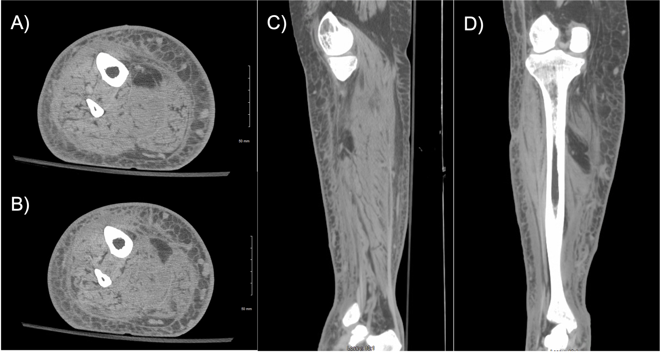
Laboratory values at that time demonstrated a white blood cell count (WBC) of 20,800cells/uL, hemoglobin 14g/dl, Erythrocyte Sedimentation Rate (ESR) of 66mm/hr, and C-Reactive Protein (CRP) of 23.8mg/dL, Glucose of 80mg/dL, sodium of 129mmol/L, and creatinine of 1.10 mg/dL. With these initial laboratory values, her Laboratory Risk Indicator for Necrotizing Fasciitis (LRINEC) score was 8, demonstrating a 93.4% positive predictive value for necrotizing fasciitis from an objective view, which also correlated with her clinical findings.19 Imaging included radiographs of the right tibia/fibula and computer topography (CT) of the right lower extremity, which demonstrated diffuse soft tissue swelling and a 0.7 cm ulceration involving the anterior/medial aspect of the mid lower extremity. A 0.6 x 4.4 x 4.5 cm collection in the subcutaneous tissues deep to the ulcer, which may represent phlegmon and/or abscess, and a 3.0 x 1.5 x 15.0 cm fluid collection in the posterior compartment of the lower extremity extending proximally to the mid-calf (Image 1 & 2).

Given the extent of soft-tissue involvement, a multistage surgical approach was selected over an amputation of the lower extremity after shared decision-making with the patient. Risk and benefits were discussed with the patient which included the need for multiple surgeries, longer hospital stays, increase in costs, and increase risk of infection compared to limb removal. Initial surgery was a fasciotomy, irrigation, debridement, and wound vacuum application followed by multiple irrigations, debridement, and wound vacuum applications surgeries. The last surgeries included irrigation, debridement, and application of a biodegradable temporizing matrix and wound vacuum.
Surgical Technique: Initial Fasciotomy, Irrigation, Debridement, and Wound Vacuum Application
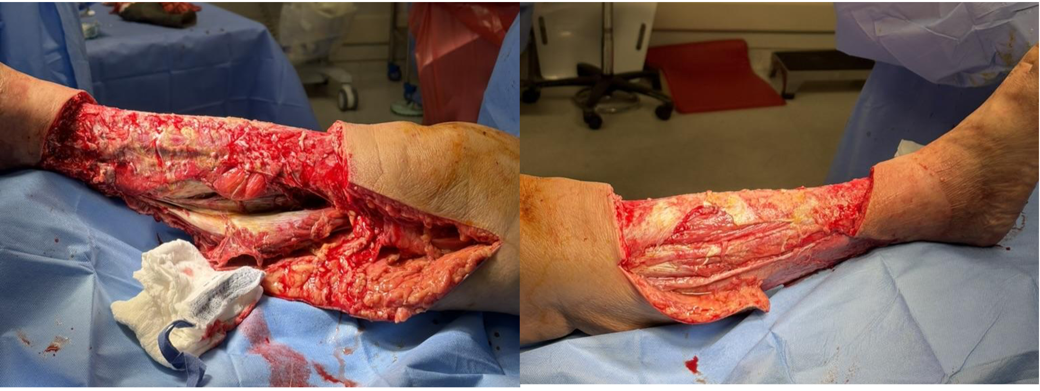
The patient had a lateral puncture wound and an active draining sinus over the medial lower leg. A medial incision incorporated the active draining sinus that was a curvilinear longitudinal incision to maximize skin bridges. Immediate thick purulent fluid erupted from the subcutaneous tissue. It tracked all the way to the level of the knee; therefore, the incision was extended medially up the thigh to the level of good, healthy tissue. Multiple cultures were obtained at this time. Betadine solution was then irrigated through the wound. A fasciotomy was performed on the lower leg, which opened the superficial posterior compartment, and immediately, thick, purulent fluid erupted again. The muscle was initially gray, but by the end of the procedure, it had turned pink and was contractile. We opened the deep posterior compartment as well and did not find any necrotic muscle. Next, an incision for the anterior and lateral compartments was made to open the fascia. There was a communication of the anterior compartment directly deep to the posterior compartment. The muscle was originally gray, but color and contractility returned after debridement. Multiple cultures were obtained again. Betadine solution was irrigated through the wound, followed by copious irrigation with 6 L of saline under gravity flow. Fresh drapes and gloves were applied. We perform a formal debridement on the right lower leg, including the skin, subcutaneous tissue, and muscle. The patient had circumferential skin loss of the lower leg between the patella and the ankle (Image 3). The lack of overlying coverage required the application of two wound vacuums. The skin was approximated with 2-0 Nylon, and then the medial wound vacuum was placed using black foam measuring 60 x 5 x 2 cm with non-disposable goods. The lateral wound measured 30 x 5 x 2 cm. Again, 2-0 Nylon was used to approximate the skin, and black foam was applied. Wound vacuums were initiated at 125 mmHg with continuous suction and an appropriate seal. An elastic compression wrap was applied to the right lower extremity to minimize swelling. Multiple cultures from this initial surgery would subsequently grow Streptococcus pyogenes (group A). The patient was initially covered with ceftriaxone, vancomycin, and clindamycin, and then transitioned to linezolid 600mg PO BID for specific coverage of S. pyogenes and its anti-toxin effect.
Surgical Technique: Irrigation, Debridement, and Application of Biodegradable Temporizing Matrix and Wound Vacuum
After three repeat irrigations, debridements, and wound vacuum applications over one week, the patient was scheduled for a repeat irrigation, debridement, and application of a biodegradable temporizing matrix. The previous wound vacuum was removed. There was exposed tibia with granulation tissue from the wound vacuums circumferentially around the leg. The tissue was pink and healthy. The right lower extremity was then prepped and draped in a sterile fashion. Cultures were obtained from the wound. At this point, the skin, subcutaneous tissue, and muscles of the right lower extremity were debrided with a knife. Betadine solution was irrigated into the wound, followed by copious irrigation with 6 L of saline at fast gravity flow. Fresh drapes and gloves were applied. The medial thigh incision could be easily closed. The subcutaneous tissue was closed with 1. Vicryl and 2-0 Vicryl. Skin was closed with 3-0 nylon. This incision measured 15 cm. This was a delayed secondary closure of the right knee wound. The remaining open wound measured 60 x 30 by 2 cm on her right lower leg with full exposed bone, tendon, and muscle. Six 10 cm x 5cm biodegradable temporizing matrices (PolyNovo, NovoSorb BTM, PolyNovo North America LLC, USA) were placed circumferentially around the lower leg and held in place by staples. We then applied adaptic, black foam, and a wound vacuum measuring 60 x 30 x 2 cm, and set the pressure to 125 mmHg with good suction and seal.
Clinical Results
At eight weeks postoperatively, the patient was ambulating without difficulty. The patient had circumferential healing wounds from the proximal⅓ of her right lower extremity to just above her ankle. The wound was well profuse and bleeding. The biodegradable temporizing matrix was adhered with granulation tissue formation but demonstrated areas where granulation tissue did not form. The proximal medial incision was closed, dry, and intact with scarring. The patient was neurovascularly intact with palpable pedal pulses. Due to a break in granulation located on the proximal aspect of the wound, a shared decision was made to perform repeat incision, debridement, and repeat application of a biodegradable temporizing matrix.
Surgical Technique: Repeat Irrigation, Debridement, and Repeat Application of Biodegradable Temporizing Matrix and Wound Vacuum
The entire area of granulating skin was very healthy and easily bled (Image 4). This was thoroughly scrubbed, and cultures were obtained to verify that no further bacterial growth occurred. The area measured 30 x 20 cm.

It was then copiously irrigated with saline and Betadine, and then saline again using gravity flow. Fresh drapes and gloves were applied. Repeat application of biodegradable temporizing matrices (PolyNovo, NovoSorb BTM, PolyNovo North America LLC, USA) was then applied, totaling six 10cm x 5cm sheets, to circumferentially wrap around the lower leg and cover all the granulation tissue. Staples were used to hold the biodegradable temporizing matrices in place (Image 5). Adaptic, black foam, and wound vacuum were then applied and set to 125 mmHg, with good suction and seal.

Clinical Results
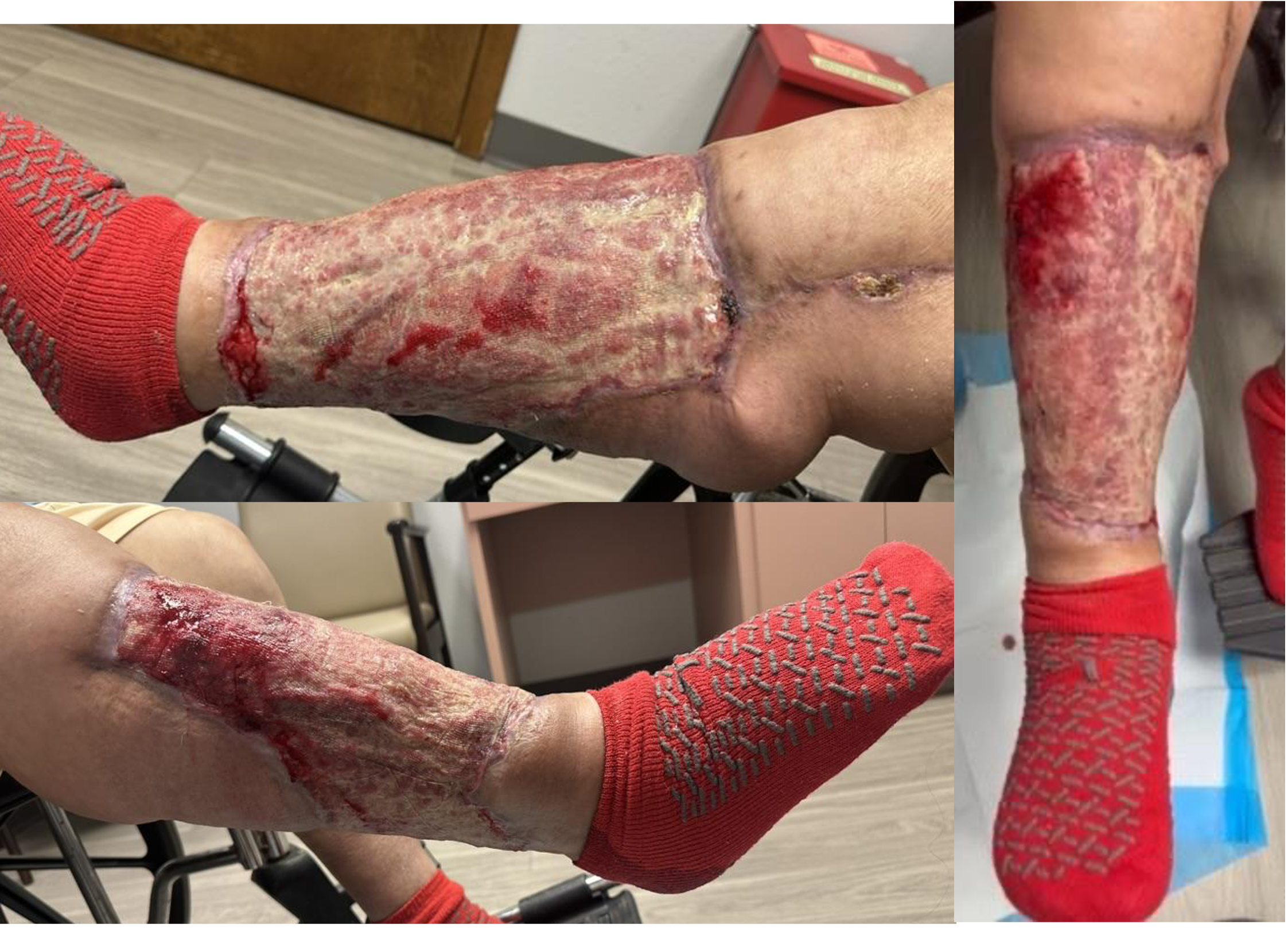
The patient’s postoperative course was unremarkable. Three months after the second application of biodegradable temporizing matrices, a physical exam demonstrated circumferential granulation tissue present about the right lower extremity with no evidence of active bleeding, drainage, or purulent material (image 6). The biodegradable temporizing matrix was adhered with granulation tissue formation. The proximal medial incision was closed, dry, and intact with scarring. The patient was neurovascularly intact with palpable pedal pulses.
Timeline
Our patient had a total of six surgeries. She was originally taken to the operating room on hospital admission day 1 for initially fasciotomy, irrigation, debridement, and application of wound vacuum. After which she had repeat irrigations, debridements, and wound vacuum applications on hospital admission days 3, 5, and 8. She then went for irrigation, debridement, and application of biodegradable temporizing matrix (BTM) and wound vacuum on day 10. She was monitored with wound vacuum in place for 5 days along with working with physical therapy. She was discharged from the hospital on hospital admission day 18. Patient then followed up with our outpatient clinic at the 3-week mark, 6-week mark, and 3-month mark after discharge. At this point a patient-physician conversations was held to talk about a repeat application of BTM due to some areas that have not fully formed granulation tissue. The patient wanted all procedures to help regrow soft tissue coverage and minimize the risk of reinfection/amputation. Patient was then readmitted to the hospital for repeat irrigation, debridement, and application of biodegradable temporizing matrix (BTM) and wound vacuum #2. Wound vaccum was kept on for 5 days and the patient worked with physical therapy. She was discharged again 1 week later and then followed up in our outpatient clinic at the 3-week, 6-week, and 3-month period.
Discussion
Necrotizing fasciitis of the lower extremity, particularly when deep multiple compartments are involved, is a limb-threatening condition associated with inherent challenges for both infection control and tissue reconstruction. In settings with delayed clinical presentation, the extent of necrotic soft-tissue development is further exacerbated, often requiring multiple irrigation and debridement procedures to achieve adequate source control. The substantial amount of tissue lost during this aggressive debridement results in large, complex wounds with exposed musculoskeletal structures, significantly complicating efforts to achieve limb salvage and soft-tissue coverage.
In patients with advanced arterial disease, flap reconstruction offers advantages and disadvantages that depend on the extent of the disease. A retrospective study used angiography to analyze blood flow and determine whether the flap pedicle would receive direct in-line or indirect flow.10 The authors demonstrated that local flap reconstruction can achieve optimal outcomes in patients with compromised arterial blood flow, no matter the severity. However, if indirect blood flow to the flap pedicle was noted, it demonstrated an increase in morbidity and higher rates of contralateral amputation.10 Additionally, another retrospective study compared the outcomes of fasciocutaneous flaps versus muscle-free flaps in patients with peripheral vascular disease.4 The authors reported higher postoperative complications and an increased need for re-intervention in the fasciocutaneous flap group compared to the muscle flap group.4 Muscle flaps may offer physiologic advantages with lower vascular resistance and more robust perfusion for wound healing in patients with significant peripheral vascular disease. Collectively, these studies suggest that free flaps may be effective in vasculopathic limbs; however, careful consideration of flap selection, preoperative vascular assessment, and medical optimization is essential to reduce complications in this patient population.5
A variety of techniques are used to achieve effective soft-tissue defect coverage following surgical resection or aggressive debridement, including traditional free flap reconstruction and biological dermal wound matrices. A retrospective analysis evaluated the use of bilayer wound matrix compared to free flap reconstruction in patients with complex scalp wounds.13 The authors concluded that local tissue rearrangement is superior for wounds measuring less than 100 cm2; however, the bilayer wound matrix achieved comparable outcomes to free flap reconstruction for larger wound defects (150-200 cm2). In addition, the use of a bilayer wound matrix was associated with shorter operative times and shorter hospital stays and may be more cost-effective.13 These findings suggest that biological matrices can serve as a viable alternative in select patients, depending on the size of the wound defect, donor-site morbidity, and resource limitations that would increase operative risks in free flap reconstruction. Free flap reconstruction remains the gold standard due to its extensive vascular coverage and utility in complex wounds; however, biological substitutes are a viable alternative for appropriately selected soft-tissue defects.
Skin substitutes, such as biodegradable temporizing matrices, come in many forms and remain a fundamental technique for treating wound defects. Skin substitutes can be derived biologically, synthetically, or biosynthetically to provide appropriate scaffolding, a growth environment, and cellular optimization for the treatment of chronic or acute wounds. There are at least 76 commercially available skin substitutes as of 2020, all of which are considered to be biodegradable and temporary.3
The most commonly used substitutes are acellular dermal matrices (e.g., AlloDerm, DermACELL, GraftJacket), derived from human or animal tissue.1,3,17 This substitute offers a decellularized scaffolding allowing host cells to infiltrate and facilitate wound healing in a growth-promoting environment. Allograft or xenograft dermal substitutes remain the most commonly commercially available acellular skin substitutes due to the long shelf life and increased supply.3 Human placental membrane grafts (e.g., EpiFix, AmnioBand) demonstrated an accelerated rate of healing in patients with chronic or burn wounds. There are composite substitutes that consist of bilayered matrix (e.g., Integra Dermal Regeneration Template, Polynovo BTM) that provide the extracellular scaffolding as well as promote the wound bed for vascularization and dermal regeneration.1,3,17 While each of these substitutes has advantages and disadvantages with respect to wound healing and outcomes, careful consideration must be given to the varied biological composition, cost, extent of wound defect, and availability.
In the presented case, BTM was used following serial debridement to resolve the active infection, stabilize the wound bed, and promote controlled tissue regeneration. The application of this matrix covered exposed musculoskeletal structures while promoting progressive neoderm formation by creating a favorable biological environment. Given the extent of tissue loss, compromised local vasculature, and continuous wound monitoring, the BTM approach was beneficial in a setting of multicompartment necrotizing infection. Continuation of care is still needed for our patient. At the 3-month mark she has circumferential granulation tissue formation but has not completely healed yet. She will continue to receive daily dressing changes with oil-based dressings (adaptic), 4x4s, abdominals bads, and finally an elastic compression bandage until wound fully heals. Patient is to follow-up in our outpatient office every month for wound evaluation and clinical checkup.
Given the medical-surgical emergency associated with this deep, multicompartment infection of NF, prioritizing compromised tissue and infection control were the primary management steps. The standard care of treatment follows aggressive debridement with surgical removal of nonviable tissue, as incomplete infection eradication may require additional unnecessary procedures.11,20 A multistage approach was used, involving multiple debridement procedures to optimize tissue conditions and progress toward more definitive coverage. This strategy allowed for a careful and controlled assessment of wound stability through multiple samples being collected for microbiological examination, and to confirm that the debridement was thorough before tissue reconstruction.
Conclusion
Management of necrotizing fasciitis in the setting of deep infections with multicompartment involvement remains complex, particularly in patients with delayed presentation, which allows the disease process to propagate. Accurate preoperative quantification of the infected area, thoughtful wound management, and careful consideration of surgical debridement are critical to optimize outcomes. This case demonstrates that a multistage strategy, combined with a biodegradable temporizing matrix, can restore soft-tissue coverage and function. This approach provides treatment of the infection without recurrent wound nonhealing. To our knowledge, this is the first report of a multistage surgical procedure for necrotizing fasciitis with extensive circumferential soft-tissue loss and the use of a biodegradable temporizing matrix, offering a potential treatment framework for similarly challenging presentations.
Author contributions: All authors provided substantial contributions to conception and design, acquisition of data, or analysis and interpretation of data; all authors drafted the article or revised it critically for important intellectual content; all authors gave final approval for the version of the article to be published; and all authors agree to be accountable for all aspects of the work in ensuring that questions related to the accuracy or integrity of any part of the work are appropriately investigated and resolved.
Level of Evidence: IV Case Report
Disclosures: There were no competing interests in this study or study design. This research received no external funding or competitive funding. No Artificial Intelligence (AI) software or writing tools were used in the research process of this study.
Disclaimer: This research was supported (in whole or in part) by HCA Healthcare and/or an HCA Healthcare affiliated entity. The views expressed in this publication represent those of the author(s) and do not necessarily represent the official views of HCA Healthcare or any of its affiliated entities.
Data Availability Statement: Data is contained within the article.
Number of Abstract Words: 380
Number of Text Words: 2996
Number of References: 20
Number of Tables/Figures: 6
Number of Videos: 0
Co-Authors
Contributing Authors/Acknowledgment: we would like to thank *** and *** for their assistance in data collection, project management, and patient presentation.
References
- Alrubaiy L, Al-Rubaiy KK. Skin Substitutes: A Brief Review of Types and Clinical Applications. Oman Med J. 2009;24(1):4-6. doi:10.5001/omj.2009.2
- Barreiro D, Mendes M, Machado P, Oliveira I, Horta R. Necrotizing fasciitis: act fast and act together. Ann Burns Fire Disasters. 2025;38(1):80-85.
- Bay C, Chizmar Z, Reece EM, et al. Comparison of Skin Substitutes for Acute and Chronic Wound Management. Semin Plast Surg. 2021;35(3):171-180. doi:10.1055/s-0041-1731463
- Bovill JD, Sayyed AA, Huffman SS, et al. A comparative analysis of fasciocutaneous versus muscle free flaps in peripheral vascular disease patients. Microsurgery. 2023;43(3):253-260. doi:10.1002/micr.31008
- Chen ACY, Lin TW, Chang KC, Chang DH. Strategic Use of Biodegradable Temporizing Matrix (BTM) in Wound Healing: A Case Series in Asian Patients. J Funct Biomater. 2024;15(5):136. doi:10.3390/jfb15050136
- Chen LL, FASOLKA B, TREACY C. Necrotizing fasciitis: A comprehensive review. Nursing (Lond). 2020;50(9):34-40. doi:10.1097/01.NURSE.0000694752.85118.62
- Hakkarainen TW, Kopari NM, Pham TN, Evans HL. Necrotizing soft tissue infections: Review and current concepts in treatment, systems of care, and outcomes. Curr Probl Surg. 2014;51(8):344-362. doi:10.1067/j.cpsurg.2014.06.001
- Li A, Dearman BL, Crompton KE, Moore TG, Greenwood JE. Evaluation of a novel biodegradable polymer for the generation of a dermal matrix. J Burn Care Res Off Publ Am Burn Assoc. 2009;30(4):717-728. doi:10.1097/BCR.0b013e3181abffca
- Li H, Lim P, Stanley E, et al. Experience with NovoSorb® Biodegradable Temporising Matrix in reconstruction of complex wounds. ANZ J Surg. 2021;91(9):1744-1750. doi:10.1111/ans.16936
- Li KR, Rohrich RN, Lava CX, Akbari CM, Attinger CE. Optimizing Lower Extremity Local Flap Reconstruction in Peripheral Vascular Disease. Ann Plast Surg. 2024;93(4):488-495. doi:10.1097/SAP.0000000000004105
- Manna B, Nahirniak P, Morrison CA. Wound Debridement. In: StatPearls. StatPearls Publishing; 2025. Accessed February 15, 2026. http://www.ncbi.nlm.nih.gov/books/NBK507882/
- McDermott J, Kao LS, Keeley JA, Grigorian A, Neville A, de Virgilio C. Necrotizing Soft Tissue Infections: A Review. JAMA Surg. 2024;159(11):1308-1315. doi:10.1001/jamasurg.2024.3365
- Othman S, Shakir S, Azoury SC, et al. Utility of Dermal Wound Matrices Compared with Local-Tissue Rearrangement and Free Flap Reconstruction for Oncologic Scalp Wounds: A Multidisciplinary Dual Matched-Pair Analysis. Plast Reconstr Surg. 2022;149(2):469-480. doi:10.1097/PRS.0000000000008774
- Schlottmann F, Obed D, Bingöl AS, März V, Vogt PM, Krezdorn N. Treatment of Complex Wounds with NovoSorb® Biodegradable Temporising Matrix (BTM)—A Retrospective Analysis of Clinical Outcomes. J Pers Med. 2022;12(12):2002. doi:10.3390/jpm12122002
- Struble SL, Patel NK, Graham EM, et al. Outcomes of Biodegradable Temporizing Matrix for Soft Tissue Reconstruction of the Hand and Extremities. Plast Reconstr Surg Glob Open. 2024;12(7):e5956. doi:10.1097/GOX.0000000000005956
- Tedesco S, Di Grezia M, Tropeano G, Altieri G, Brisinda G. Necrotizing soft tissue infections: a surgical narrative review. Updat Surg. 2025;77(4):1239-1251. doi:10.1007/s13304-025-02222-0
- Vecin NM, Kirsner RS. Skin substitutes as treatment for chronic wounds: current and future directions. Front Med. 2023;10:1154567. doi:10.3389/fmed.2023.1154567
- Wallace HA, Perera TB. Necrotizing Fasciitis. In: StatPearls. StatPearls Publishing; 2025. Accessed February 15, 2026. http://www.ncbi.nlm.nih.gov/books/NBK430756/
- Wong CH, Khin LW, Heng KS, Tan KC, Low CO. The LRINEC (Laboratory Risk Indicator for Necrotizing Fasciitis) score: a tool for distinguishing necrotizing fasciitis from other soft tissue infections. Crit Care Med. 2004;32(7):1535-1541. doi:10.1097/01.ccm.0000129486.35458.7d
- Zhou L, Li H, Luo G, CANF experts group representing the Chinese Burn Association, Editorial Board of the Chinese Journal of Burns and Wounds, Burn Medicine Branch of China International Exchange and Promotion Association for Medical and Healthcare. Consensus on the Diagnosis and Treatment of Adult Necrotizing Fasciitis (2025 Edition). Burns Trauma. 2025;13:tkaf031. doi:10.1093/burnst/tkaf031
Author contributions
All authors provided substantial contributions to conception and design, acquisition of data, or analysis and interpretation of data; all authors drafted the article or revised it critically for important intellectual content; all authors gave final approval for the version of the article to be published; and all authors agree to be accountable for all aspects of the work in ensuring that questions related to the accuracy or integrity of any part of the work are appropriately investigated and resolved.
Level of Evidence
IV Case Report
Disclosures
There were no competing interests in this study or study design. This research received no external funding or competitive funding. No Artificial Intelligence (AI) software or writing tools were used in the research process of this study.
Disclaimer
This research was supported (in whole or in part) by HCA Healthcare and/or an HCA Healthcare affiliated entity. The views expressed in this publication represent those of the author(s) and do not necessarily represent the official views of HCA Healthcare or any of its affiliated entities.
Data Availability Statement
Data is contained within the article.
- Number of Abstract Words: 380
- Number of Text Words: 2996
- Number of References: 20
- Number of Tables/Figures: 6
- Number of Videos: 0
Keywords: Necrotizing Fasciitis, Application of Skin Graft, Biodegradable Temporizing Matrix, Complex Lower Extremity Wound, Multi-Compartment Involvement
Co-Authors: Contributing Authors/Acknowledgment: we would like to thank *** and *** for their assistance in data collection, project management, and patient presentation.