Correction notice:
This article was corrected on September 28, 2026, to add Nikolas Vukotic, Kansas City University College of Osteopathic Medicine, Kansas City, Missouri, USA, to the author list. The author’s affiliation and contribution information have also been updated. https://aoao.org/2026/09/11/a-correction-has-been-published/
Curtis Wilkerson¹; Sebastian Laguna²; Nikolas Vukotic³; Jamil Haddad⁴; Shafic Sraj⁵
¹ Michigan State University College of Osteopathic Medicine, East Lansing, Michigan, USA
² Midwestern University Arizona College of Osteopathic Medicine, Glendale, Arizona, USA
³ Kansas City University College of Osteopathic Medicine, Kansas City, Missouri, USA
⁴ McLaren Macomb Hospital, Mount Clemens, Michigan, USA
⁵ Department of Orthopaedics, West Virginia University School of Medicine, Morgantown, West Virginia, USA
Introduction / Objective
Acute calcific periarthritis (ACP) is an uncommon inflammatory condition caused by the deposition of dystrophic calcium hydroxyapatite crystals in periarticular soft tissues. While it typically affects larger joints such as the shoulder or wrist, its occurrence in the digits is rare and frequently misdiagnosed as infection, gout, or osteoarthritis. The objective of this report is to highlight the importance of considering ACP in the differential diagnosis of isolated digital joint pain and to outline effective diagnostic and treatment strategies.
Methods
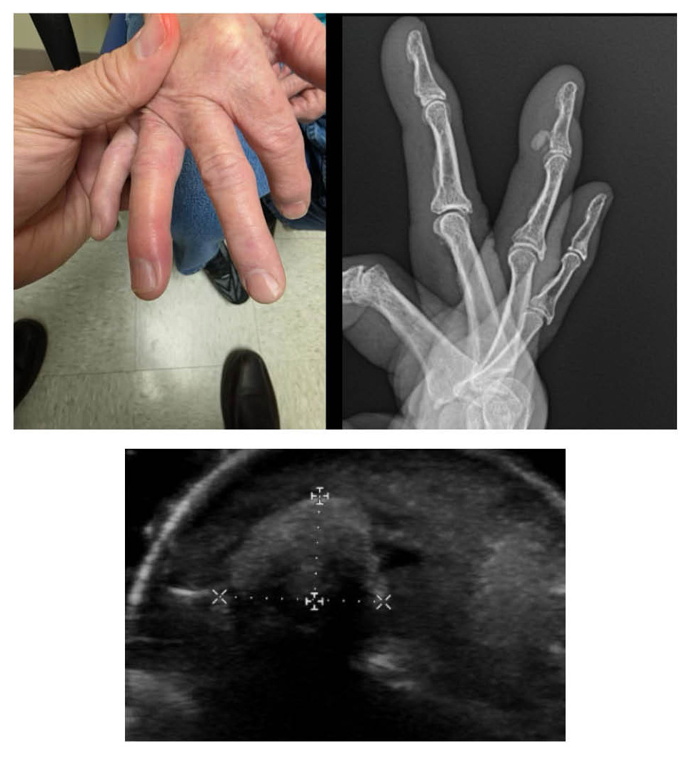
We present the case of a 66-year-old female who reported progressive pain in the right ring finger over a 2–3-month period. Clinical examination revealed localized swelling, severe tenderness, limited range of motion, and pain rated 10/10. Initial working diagnosis was osteoarthritis. However, imaging studies, including plain radiographs and musculoskeletal ultrasound, were performed to further evaluate the cause of symptoms.
Results
Radiographs demonstrated a well-circumscribed radiopaque deposit adjacent to the distal phalanx. Ultrasound imaging confirmed the presence of a hyperechoic calcific deposit without evidence of flexor tendon involvement. Surgical incision and drainage were performed, and liquified calcific material was excised. Pathologic evaluation confirmed dystrophic calcification consistent with ACP. The patient experienced complete resolution of symptoms following normal postoperative surgical course with no residual pain.
Conclusions
This case emphasizes the diagnostic value of imaging modalities in identifying ACP in atypical locations such as the digits. Early recognition can prevent misdiagnosis and unnecessary interventions. Given the rarity of digital ACP, this case adds to the limited literature and reinforces the need for heightened clinical suspicion when typical diagnoses do not align with imaging or clinical progression.